. 2000 Dec 15;19(24):6792-803.
doi: 10.1093/emboj/19.24.6792.

P/CAF-mediated acetylation regulates the function of the basic helix-loop-helix transcription factor TAL1/SCL

Affiliations

P/CAF-mediated acetylation regulates the function of the basic helix-loop-helix transcription factor TAL1/SCL

S Huang et al. EMBO J. .

Abstract

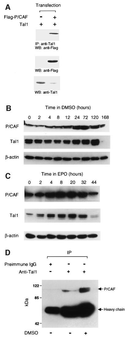
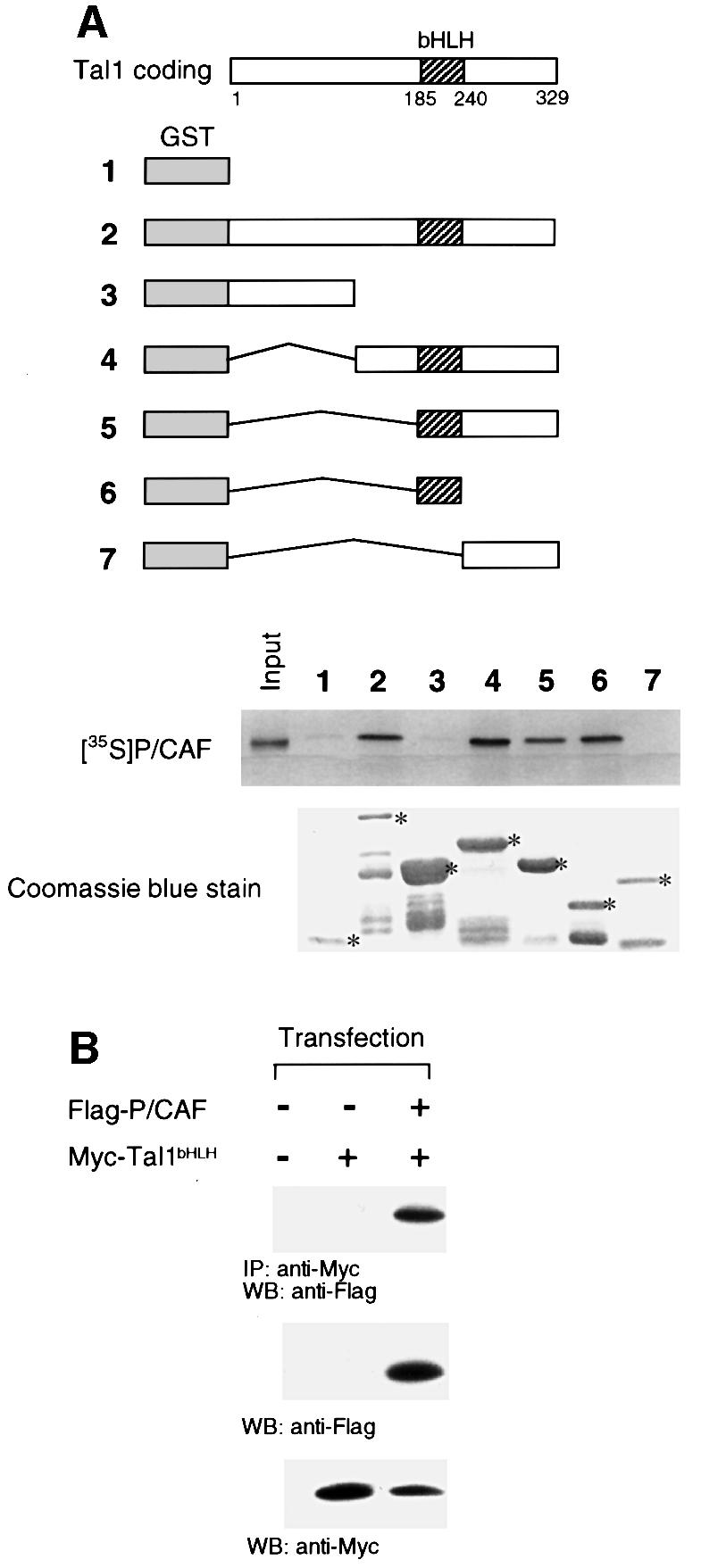
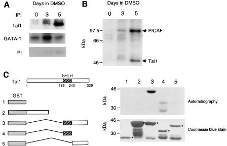
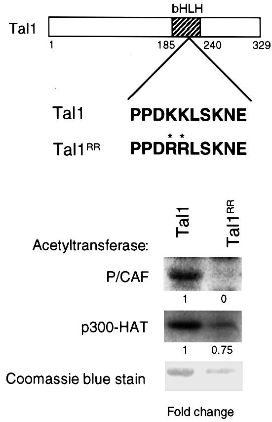
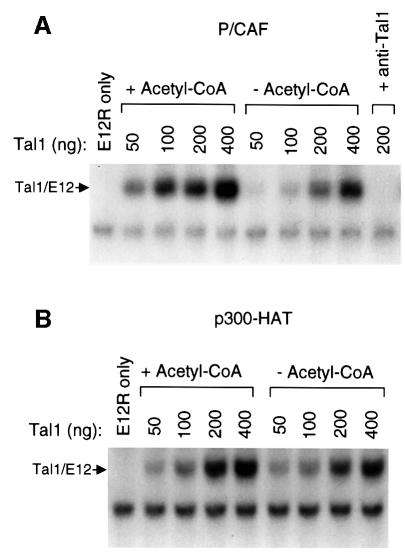
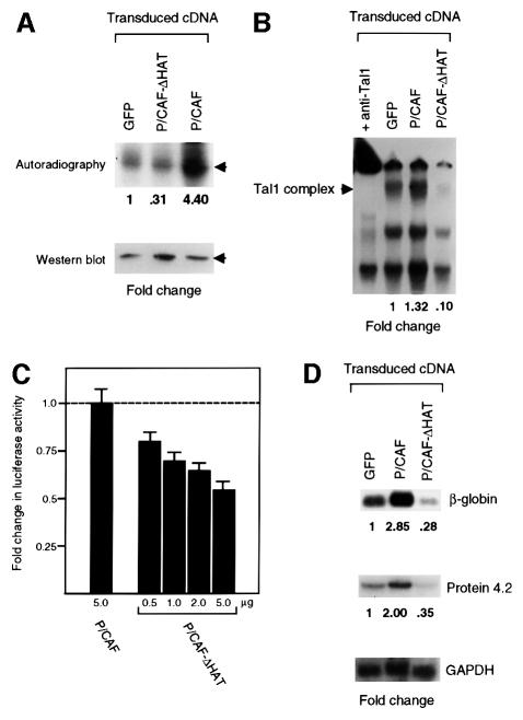
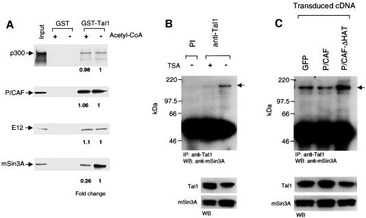
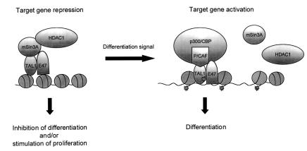
The basic helix-loop-helix transcription factor TAL1 (or SCL) is a critical regulator of hematopoietic and vascular development and is misexpressed in the majority of patients with T-cell acute lymphoblastic leukemia. We found previously that TAL1 could interact with transcriptional co-activator and co-repressor complexes possessing histone acetyltransferase and deacetylase activities, respectively. Here, we report that TAL1 is subject to acetylation in vivo and can be acetylated by p300 and the p300/CBP-associated factor P/CAF in vitro. P/CAF-mediated acetylation, which mapped to a lysine-rich motif in the loop region, increased TAL1 binding to DNA while selectively inhibiting its interaction with the transcriptional co-repressor mSin3A. Furthermore, P/CAF protein, TAL1-P/CAF interaction and TAL1 acetylation increased significantly in murine erythroleukemia cells induced to differentiate in culture, while enforced expression of an acetylation-defective P/CAF mutant inhibited endogenous TAL1 acetylation, TAL1 DNA-binding activity, TAL1-directed transcription and terminal differentiation of these cells. These results reveal a novel mechanism by which TAL1 activity is regulated and implicate acetylation of this transcription factor in promotion of erythroid differentiation.


Figures

None
Fig. 1. P/CAF co-immunoprecipitates with TAL1 in cellular extracts. (A) Co-immunoprecipitation of P/CAF with TAL1 in transfected HeLa cells. HeLa cells were transfected with expression vectors for TAL1 or Flag-tagged P/CAF, cellular extracts immunoprecipitated with TAL1 antibody, and immunoprecipitates subjected to western blot (WB) analysis with a Flag epitope-specific antibody. P/CAF protein was identified only in extracts from TAL1- and P/CAF-co-transfected cells (top). Direct western blot analysis confirmed that both P/CAF (middle) and TAL1 (bottom) proteins were expressed in transfected cells. (B) Increase in P/CAF and TAL1 expression in differentiating MEL cells. Lysates of MEL cells incubated with 1.8% DMSO for the indicated number of hours were fractionated by SDS–PAGE and transferred to a PVDF membrane, which was incubated sequentially with antibodies to P/CAF (top), TAL1 (middle) and β-actin (bottom). Antibody binding was visualized by enhanced chemiluminescence. Multiple forms of TAL1 are evident, reflecting phosphorylation, alternative translational initiation (‘leaky’ ribosomal scanning), acetylation or any combination of these. (C) Increase in P/CAF and TAL1 expression in Epo-stimulated FVA cells. Lysates of FVA cells incubated with Epo for the indicated number of hours were subjected to immunoblot analysis as described above. (D) Co-immunoprecipitation of P/CAF with TAL1 in extracts from differentiating MEL cells. Cellular extracts from 2 × 10⁶ MEL cells cultured for 5 days with (+) or without (–) 1.8% DMSO were incubated with TAL1 pre-immune IgG or Tal1 antibody and the resulting immunoprecipitates subjected to western blot analysis with P/CAF-specific antibody.
None
Fig. 2. The bHLH domain of TAL1 is necessary and sufficient for its interaction with P/CAF. (A) Schematic representation of GST–TAL1 fusion proteins used in GST pull-down assays. Constructs 2–7 contain amino acids 1–329, 1–144, 142–329, 185–329, 185–240 and 242–329, respectively, of the TAL1 coding sequence. 35S-labeled P/CAF was incubated with GST or GST–TAL1 proteins pre-adsorbed to glutathione–Sepharose beads. Specifically bound P/CAF was eluted from washed beads and visualized by fluorography following SDS–PAGE. Input represents 10% of the in vitro translated P/CAF protein used in the assay. The relative amounts of fusion proteins used (denoted by asterisks) are shown in the accompanying photograph of a Coomassie Blue-stained SDS–polyacrylamide gel. (B) HeLa cells were transfected with expression vectors for Myc-tagged TAL1 bHLH polypeptide (Myc-Tal1bHLH) or Flag-tagged P/CAF (Flag-P/CAF), cellular extracts immunoprecipitated (IP) with antibody to Myc epitope, and immunoprecipitates subjected to western blot (WB) analysis with antibody to the Flag epitope. P/CAF protein was identified only in extracts from TAL1 bHLH- and P/CAF-co-transfected cells (top). Direct western blot (WB) analysis confirmed that both P/CAF (middle) and TAL1 (bottom) were expressed in transfected cells.
None
Fig. 3. TAL1 is acetylated by P/CAF in vitro and in vivo. (A) TAL1 acetylation in vivo. MEL cells treated with DMSO for the indicated numberof days were pulse-labeled with [3H]acetate in the presence of 50 nM TSA and lysed. Lysates were subjected to immunoprecipitation with TAL1 antibody (top), GATA-1 antibody (middle) or TAL1 pre-immune IgG (bottom). The resulting immunoprecipitates were fractionated by SDS–PAGE and analyzed for incorporation of [3H]acetate by autoradiography. (B) Replicate experiment of the above in which MEL cells were labeled with [3H]acetate and cellular extracts immunoprecipitated with TAL1 antibody. In addition to TAL1, a radiolabeled protein with the mobility expected for P/CAF was precipitated, consistent with P/CAF autoacetylation. (C) P/CAF acetylation of TAL1 in vitro. Purified GST (1) and GST fusion proteins containing amino acids 1–144 (2), 185–329 (3), 185–240 (4) and 242–329 (5) of the TAL1 coding sequence were incubated with [14C]acetyl-CoA and baculovirus-expressed P/CAF. Reaction products were fractionated by SDS–PAGE and the dried gel exposed to X-ray film. The relative amounts of fusion proteins used (denoted by asterisks) are shown in the accompanying photograph of a Coomassie Blue-stained SDS–polyacrylamide gel.
None
Fig. 4. TAL1 is differentially acetylated by P/CAF and p300. Purified GST fusion protein containing the complete TAL1 coding region (Tal1) and the corresponding fusion protein with arginine substitutions (K221R K222R) for the indicated lysine residues (denoted by asterisks) in the loop region of the bHLH domain (Tal1RR) were used as substrates in an in vitro acetylation assay. Baculovirally expressed P/CAF (top) or bacterially expressed p300-HAT (middle) was incubated with these fusion proteins and [14C]acetyl-CoA, and the reaction products were fractionated by SDS–PAGE and analyzed by autoradiography. The relative amount of acetylation (fold change) was adjusted for differences in fusion protein concentration as determined by Coomassie Blue staining of the gel (bottom).
None
Fig. 5. TAL1 DNA-binding activity is augmented by P/CAF but notby p300 acetylation. (A) Purified GST–TAL1 protein in the indicated amounts and in vitro translated E12 protein (E12R) were incubated with baculovirally expressed P/CAF, with (+) or without (–) acetyl-CoA, for 1 h at 30°C. The reaction mixture was then incubated with a 32P-labeled double-stranded oligonucleotide corresponding to a preferred TAL1/E12-binding site for 30 min at room temperature, and DNA-binding activity was analyzed by EMSA. The presence of TAL1 in the binding complex marked Tal1/E12 was confirmed by the disappearance of this complex following addition of TAL1 antibody (+ anti-Tal1) to the binding reaction prior to electrophoresis. (B) Purified GST–TAL1 protein was incubated with the p300 HAT domain under acetylating and non-acetylating conditions as described in (A) and the effect of p300-stimulated acetylation on TAL1/E12 DNA binding determined by EMSA.
None
Fig. 6. P/CAF acetyltransferase activity is important for TAL1 acetylation, DNA-binding activity, transcriptional activity and erythroleukemia cell differentiation. (A) The importance of P/CAF acetyltransferase activity for TAL1 acetylation in vivo. MEL cells transduced with retroviral vectors expressing wild-type P/CAF (P/CAF), a HAT-defective P/CAF mutant (P/CAF-ΔHAT) or MSCV-IRES-GFP parental vector (GFP) were selected for GFP expression by FACS, incubated with 1.8% DMSO for 5 days, and then pulse-labeled with [3H]acetate as described in Figure 3B. Cellular extracts were subjected to immunoprecipitation with TAL1 antibody and acetylation analyzed by autoradiography. The fold increase or decrease in acetylation was related to cells transduced with empty vector (GFP) and normalized for TAL1 protein expression (bottom panel). (B) The importance of P/CAF acetyltransferase activity for TAL1 DNA-binding activity in vivo. MEL cells were transduced with the retroviral vectors described above, selected for GFP expression by FACS and incubated with 50 nM TSA for 5 h. Nuclear extracts were incubated with a 32P-labeled, double-stranded oligonucleotide corresponding to a preferred TAL1/E2A-binding site for 30 min at room temperature, and DNA-binding activity was analyzed by EMSA. The presence of TAL1 in the binding complex marked Tal1 complex was confirmed by the shift in the mobility of this complex following addition of TAL1 antibody (+ anti-Tal1) to the binding reaction prior to electrophoresis. (C) MEL cells induced to differentiate by incubation with 1.5% DMSO for 3 days were transfected with a firefly luciferase reporter linked to four copies of the GAL4 DNA-binding site and minimum TK promoter (1.0 µg), an expression vector for a GAL4–full-length TAL1 fusion protein (1.0 µg), an expression vector for either full-length P/CAF (1.0–5.0 µg) or a mutant lacking the HAT domain (0.5–5.0 µg) and an expression vector for Renilla luciferase (0.1 µg). Transfected cells and controls transfected with the reporter construct alone were returned to culture for 24 h, treated with 100 nM TSA for an additional 24 h, and lysed for measurement of luciferase activity. The luciferase activity induced by GAL4–TAL1, as corrected for reporter activity elicited by GAL41–147 and normalized for variation in transfection efficiency, was assigned a value of 1, and the fold induction or repression of reporter activity by the co-transfected P/CAF constructs was determined. Plotted is the mean fold change in luciferase activity ± SE from four independent experiments. (D) Importance of P/CAF acetyltransferase activity for DMSO-induced differentiation of MEL cells. MEL cells were transduced with the retroviral vectors described above, selected for GFP expression by FACS and incubated with 1.8% DMSO for 5 days. RNA from these cells was fractionated in a formaldehyde–agarose gel, transferred to a Nytran membrane and sequentially hybridized with radiolabeled probes for β-globin, Protein 4.2 and GAPDH. The fold change in expression of β-globin and Protein 4.2 mRNAs was determined relative to vector-transduced cells and normalized for GAPDH mRNA expression.
None
Fig. 7. Acetylation of TAL1 by P/CAF selectively impairs its interaction with mSin3A. (A) Glutathione–Sepharose beads pre-adsorbed with GST or GST–TAL11–329 fusion protein treated with purified P/CAF in the presence or absence of acetyl-CoA were mixed with [35S]methionine-labeled p300, P/CAF, E12 or mSin3A for 1 h at room temperature. Specifically bound protein was eluted from washed beads and visualized by fluorography following SDS–PAGE. The fold difference in binding of the indicated proteins was determined from image analysis of exposed X-ray film. Input represents 10% of the radiolabeled protein used in the assay. (B) Reduced association of mSin3A with hyperacetylated TAL1 protein in vivo. Lysates of MEL cells that had been treated for 5 h with (+) or without (–) 50 nM TSA were incubated with antibody to TAL1 or TAL1 pre-immune immunoglobulin (PI) and the resulting immunoprecipitates (IP) subjected to western blot analysis with an mSin3A-specific antibody. mSin3A protein that co-immunoprecipitated with TAL1 was visualized by enhanced chemiluminescence and is marked by the arrowhead. The presence of both TAL1 and mSin3A protein in these cells was verified by direct western blot (WB) analysis of extracts. (C) The importance of P/CAF acetyltransferase activity for TAL1 interaction with mSin3A in vivo. Cellular lysates were prepared from MEL cells transduced with P/CAF, P/CAF-ΔHAT or parental retrovirus and treated with 50 nM TSA for 1 h. mSin3A protein associated with TAL1 was quantitated by co-immunoprecipitation analysis as described in (B). Comparable amounts of TAL1 and mSin3A protein were present in these cells as determined by western blot (WB) analysis of extracts.
None
Fig. 8. Model of TAL1–co-regulator interactions in erythroleukemia cell differentiation. TAL1 interacts with the nuclear co-repressor mSin3A and associated histone deacetylase HDAC1 in undifferentiated MEL cells. This co-repressor complex mediates TAL1-dependent transcriptional repression and restricts the ability of these cells to respond to differentiation inducers. Addition of DMSO results in formation of TAL1–P/CAF and TAL1–p300/CBP complexes and acetylation of both TAL1 and histones. P/CAF-mediated TAL1 acetylation, in addition, decreases TAL1 interaction with mSin3A-containing complexes. In combination, these effect the conversion of TAL1 from transcriptional repressor to activator and augment cellular differentiation. Although the figure shows both P/CAF and p300/CBP present in a single complex in differentiated cells, this has not been formally established.

Similar articles

Cited by

References

    1. Akazawa C., Ishibashi,M., Shimizu,C., Nakanishi,S. and Kageyama,R. (1995) A mammalian helix–loop–helix factor structurally related to the product of Drosophila proneural gene atonal is a positive transcriptional regulator expressed in the developing nervous system. J. Biol. Chem., 270, 8730–8738. - PubMed
    1. Aplan P.D., Nakahara,K., Orkin,S.H. and Kirsch,I.R. (1992) The SCL gene product: a positive regulator of erythroid differentiation. EMBO J., 11, 4073–4081. - PMC - PubMed
    1. Arany Z., Newsome,D., Oldread,E., Livingston,D.M. and Eckner,R. (1995) A family of transcriptional adaptor proteins targeted by the E1A protein. Nature, 374, 81–84. - PubMed
    1. Atchley W.R., Wollenberg,K.R., Fitch,W.M., Terhalle,W. and Dress,A.W. (2000) Correlations among amino acid sites in bHLH protein domains: an information theoretic analysis. Mol. Biol. Evol., 17, 164–178. - PubMed
    1. Bannister A.J. and Kouzarides,T. (1995) CBP-induced stimulation of c-Fos activity is abrogated by E1A. EMBO J., 14, 4758–4762. - PMC - PubMed

Publication types

MeSH terms