. 2000 Dec 15;19(24):6860-9.
doi: 10.1093/emboj/19.24.6860.

The spliceosome deposits multiple proteins 20-24 nucleotides upstream of mRNA exon-exon junctions

Affiliations

The spliceosome deposits multiple proteins 20-24 nucleotides upstream of mRNA exon-exon junctions

H Le Hir et al. EMBO J. .

Abstract

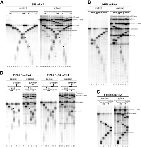
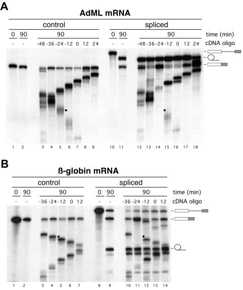
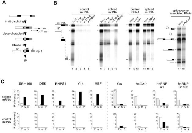
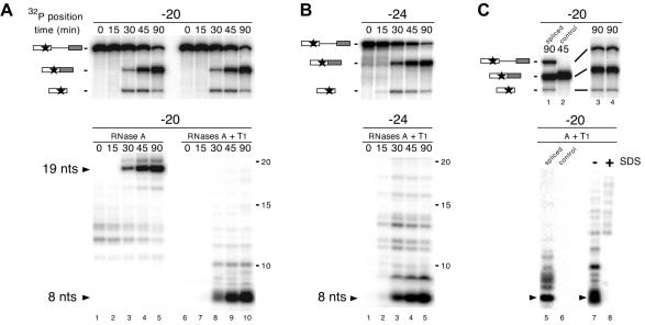
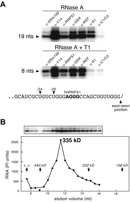
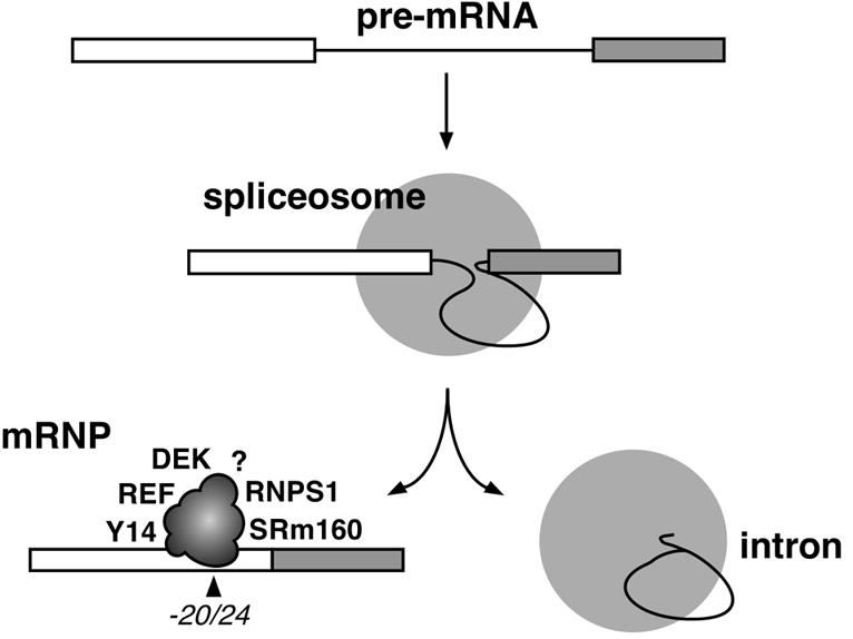
Eukaryotic mRNAs exist in vivo as ribonucleoprotein particles (mRNPs). The protein components of mRNPs have important functions in mRNA metabolism, including effects on subcellular localization, translational efficiency and mRNA half-life. There is accumulating evidence that pre-mRNA splicing can alter mRNP structure and thereby affect downstream mRNA metabolism. Here, we report that the spliceosome stably deposits several proteins on mRNAs, probably as a single complex of approximately 335 kDa. This complex protects 8 nucleotides of mRNA from complete RNase digestion at a conserved position 20-24 nucleotides upstream of exon-exon junctions. Splicing-dependent RNase protection of this region was observed in both HeLa cell nuclear extracts and Xenopus laevis oocyte nuclei. Immunoprecipitations revealed that five components of the complex are the splicing-associated factors SRm160, DEK and RNPS1, the mRNA-associated shuttling protein Y14 and the mRNA export factor REF. Possible functions for this complex in nucleocytoplasmic transport of spliced mRNA, as well as the nonsense-mediated mRNA decay pathway, are discussed.


Figures

None
Fig. 1. A splicing-dependent protection of mRNAs from targeted RNase H cleavage. (A) Uniformly labeled TPI control mRNA (lanes 1–15) or TPI pre-mRNA (lanes 16–30) was incubated under splicing conditions in HeLa cell nuclear extract for 0 (lanes 1 and 16), 45 (lanes 2–15) or 90 min (lanes 17–30). Aliquots of the 45 and 90 min reactions were further incubated with the cDNA oligos indicated (short bars underneath the mRNA schematic at the top; lanes 3–15 and 18–30). RNAs were then separated by 14% denaturing PAGE. Each oligo was named (e.g. –96, –24, 0, 48 in top panels) according to its center position relative to the exon–exon junction, which was defined as 0. Splicing substrates, intermediates and products are indicated on the right. Black dots indicate specific mRNA cleavage fragments that are markedly reduced with spliced mRNA compared with control mRNA. Similar RNase H analyses were performed with control and spliced (B) AdML mRNAs, (C) β-globin mRNAs and (D) PIP85.B (left) and PIP85.B+10 (right) mRNAs. The triangle indicates the same cDNA oligo, which was alternatively centered at –12 and –24 for PIP85.B and PIP85.B+10 mRNAs, respectively. Likewise, the asterisk indicates a single cDNA oligo centered at –24 and –36 for each mRNA, respectively. AdML, β-globin and PIP85.B RNAs were separated by 15, 8 and 15% denaturing PAGE, respectively.
None
Fig. 2. A splicing-dependent protection of AdML (A) and β-globin (B) mRNAs from RNase H cleavage in X.laevis oocytes. Control mRNA [(A) lanes 1–9; (B) lanes 1–7] or pre-mRNA [(A) lanes 10–18; (B) lanes 8–14] was injected into oocyte nuclei. Nuclear RNAs were collected immediately [(A) lanes 1 and 10; (B) lanes 1 and 8] or 90 min after injection [(A) lanes 2–9 and 11–18; (B) lanes 2–7 and 9–14]. Where indicated, nuclear RNA samples were incubated with individual cDNA oligos to activate endogenous RNAse H [(A) lanes 3–9 and 12–18; (B) lanes 3–7 and 10–14]. AdML and β-globin RNAs were separated by 15 and 10% denaturing PAGE, respectively. For uncleaved and cleaved samples, one and four oocyte equivalents of RNA, respectively, from pools of 10 oocytes were loaded per lane. Splicing substrates and products are indicated on the right. Black dots indicate specific mRNA cleavage fragments that are markedly reduced with spliced mRNA compared with control mRNA.
None
Fig. 3. Identification of proteins associated with the exon–exon junction region by RNA co-IP. (A) Scheme of co-IP strategy. AdML pre-mRNA was spliced for 2 h, and mRNP was separated from spliceosomes by glycerol gradient fractionation. Spliced mRNA was subsequently cleaved using RNase H and two oligos (–) to produce three fragments: 5′ (white), m (black) and 3′ (shaded). The fragment ‘m’ contains the –20/24 protected region (gray oval). This reaction mixture was then subjected to IP with different antibodies. Control mRNA was analyzed in parallel reactions except that it was not fractionated on a glycerol gradient. (B) Representative RNA co-IP experiments performed with antibodies against Y14, DEK, TAP-C, RNPS1, REF, hnRNPA1 (A1) and 3mCAP. Lanes 1, 6, 11, 14 and 17 correspond to one-fifteenth of input RNA. Lane 18 corresponds to co-IP of spliceosome-associated RNAs. AdML RNAs were analyzed as indicated in Figure 1. Structures of splicing substrates, intermediates and products as well as mRNA fragments are diagrammed. (C) Co-IP efficiencies of mRNA fragments (5′, m and 3′) and spliceosomes (S, taken as splicing intermediates) with antibodies against the species indicated. Upper and lower rows of histograms correspond to fragments of spliced and control mRNAs, respectively. Co-IP efficiencies are averages of 2–5 independent experiments.
None
Fig. 4. Complete RNase digestion of singly labeled RNAs. (A) Upper panel: splicing time courses for AdML pre-mRNAs containing a single labeled phosphate (star) positioned 20 nt upstream of the 5′ splice site. Splicing substrates, intermediates and products are indicated on the left. RNAs were analyzed as in Figure 1. Lower panel: complete RNase A (left) or RNase A + T1 (right) digestion of RNAs at each time point yielded 19 and 8 nt protected fragments (arrows). Protected fragments were separated by 20% denaturing PAGE. (B) Same as (A) with AdML pre-mRNAs containing a single labeled phosphate positioned 24 nt upstream of the 5′ splice site. (C) Upper panels: AdML pre-mRNA (lanes 1, 3 and 4) or control mRNA (lane 2) containing a single labeled phosphate positioned 20 nt upstream of the 5′ splice site was incubated under splicing conditions for the time indicated and separated by 20% (lanes 1 and 2) or 15% (lanes 3 and 4) denaturing PAGE. Lower panels: aliquots of each splicing reaction were subjected to complete RNase A + T1 digestion with (lane 8) or without (lanes 5–7) prior SDS treatment. Protected fragments were separated as in (A).
None
Fig. 5. Immunoprecipitation of the protected fragments from spliced mRNA and molecular mass determination of the –20/24 complex. (A) Co-IP of 19 nt (upper panel) or 8 nt (middle panel) protected fragments generated by RNase A or RNase A + T1 digestion, respectively, of spliced AdML mRNA containing a single labeled phosphate at position –20. Antibodies are as indicated. Lower panel: sequence of AdML mRNA exon 1 between position –31 and the exon–exon junction. Arrows and bold letters indicate –20 and –24 positions and the likely binding site of hnRNP A1, respectively. (B) Superose 6 gel filtration profile of the complex associated with the RNase A-resistant 19 nt fragment of spliced mRNA labeled at position –20 (top). The amount of labeled fragment in each fraction (bottom) was determined by PhosphorImaging (PI units). Elution positions of molecular weight standards and the void volume (v.v.) are indicated.
None
Fig. 6. Schematic illustration of the –20/24 complex deposited on mRNA as a consequence of pre-mRNA splicing. Identified proteins are indicated at arbitrarily chosen positions. The question mark symbolizes potential unidentified component(s).

References

    1. Alexiadis V., Waldmann,T., Andersen,J., Mann,M., Knippers,R. and Gruss,C. (2000) The protein encoded by the proto-oncogene DEK changes the topology of chromatin and reduces the efficiency of DNA replication in a chromatin-specific manner. Genes Dev., 14, 1308–1312. - PMC - PubMed
    1. Bachi A. et al. (2000) The C-terminal domain of TAP interacts with the nuclear pore complex and promotes export of specific CTE-bearing RNA substrates. RNA, 6, 136–158. - PMC - PubMed
    1. Bear J., Tan,W., Zolotukhin,A.S., Tabernero,C., Hudson,E.A. and Felber,B.K. (1999) Identification of novel import and export signals of human TAP, the protein that binds to the constitutive transport element of the type D retrovirus mRNAs. Mol. Cell. Biol., 19, 6306–6317. - PMC - PubMed
    1. Blencowe B.J., Nickerson,J.A., Issner,R., Penman,S. and Sharp,P.A. (1994) Association of nuclear matrix antigens with exon-containing splicing complexes. J. Cell Biol., 127, 593–607. - PMC - PubMed
    1. Blencowe B.J., Issner,R., Nickerson,J.A. and Sharp,P.A. (1998) A coactivator of pre-mRNA splicing. Genes Dev., 12, 996–1009. - PMC - PubMed

Publication types

MeSH terms