. 2000 Dec 15;19(24):6870-81.
doi: 10.1093/emboj/19.24.6870.

Molecular basis of sequence-specific recognition of pre-ribosomal RNA by nucleolin

Affiliations

Molecular basis of sequence-specific recognition of pre-ribosomal RNA by nucleolin

F H Allain et al. EMBO J. .

Abstract

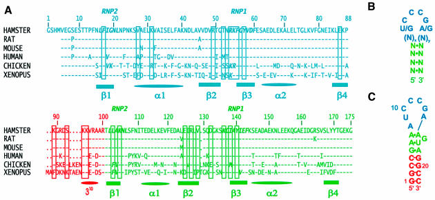
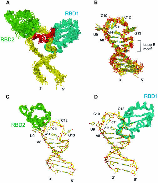
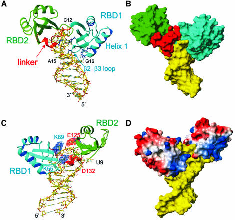
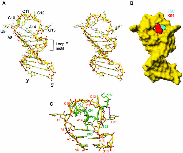
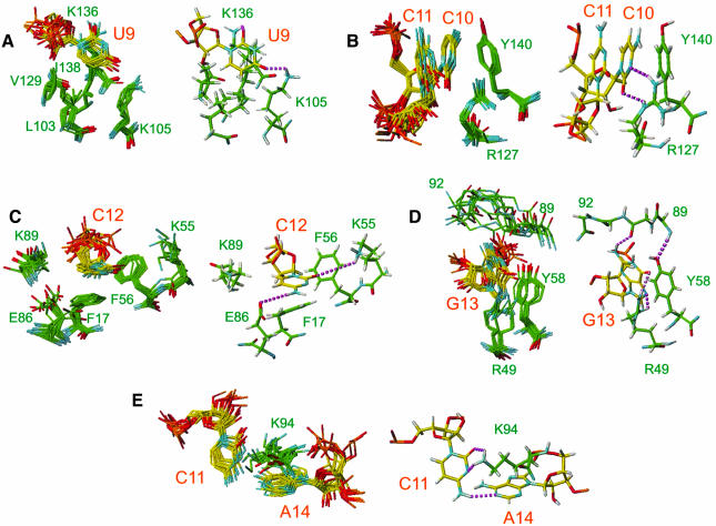
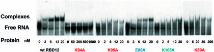
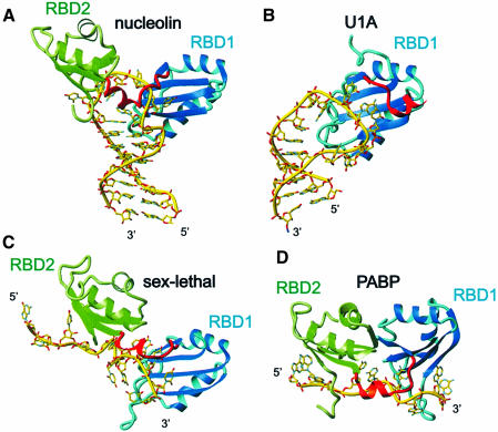
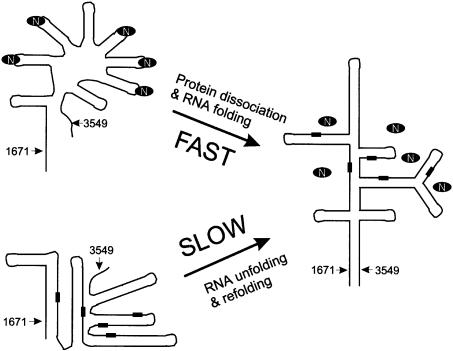
The structure of the 28 kDa complex of the first two RNA binding domains (RBDs) of nucleolin (RBD12) with an RNA stem-loop that includes the nucleolin recognition element UCCCGA in the loop was determined by NMR spectroscopy. The structure of nucleolin RBD12 with the nucleolin recognition element (NRE) reveals that the two RBDs bind on opposite sides of the RNA loop, forming a molecular clamp that brings the 5' and 3' ends of the recognition sequence close together and stabilizing the stem-loop. The specific interactions observed in the structure explain the sequence specificity for the NRE sequence. Binding studies of mutant proteins and analysis of conserved residues support the proposed interactions. The mode of interaction of the protein with the RNA and the location of the putative NRE sites suggest that nucleolin may function as an RNA chaperone to prevent improper folding of the nascent pre-rRNA.


Figures

None
Fig. 1. (A) Sequence and numbering of the hamster nucleolin RBD12 used in this study and sequence alignment [BLAST (Altschul et al., 1997)] with the nucleolin RBD12 of rat, mouse, human, chicken and Xenopus. Residues from RBD1, RBD2 and the linker are shown in cyan, green and red, respectively. The secondary structure elements are indicated below the sequences. In the sequence alignment, a dash indicates that the residue is the same as in the hamster nucleolin and a dot indicates where there is a gap relative to Xenopus, which is six residues longer than hamster nucleolin. The residues involved in either protein–RNA interactions and/or interdomain interactions in the hamster nucleolin RBD12–sNRE complex are boxed. The conserved octapeptide RNP1 and hexapeptide RNP2 characteristic of the RBD/RNP/RRM motif (Birney et al., 1993) are indicated. (B) Schematic of the consensus NRE sequence and secondary structure. (C) Sequence and secondary structure of the sNRE used in these studies. Nucleotides 3–20 are the sequence identified by in vitro selection (Ghisolfi-Nieto et al., 1996).
None
Fig. 2. Superpositions of the ensemble of the 19 lowest energy structures of the nucleolin RBD12–sNRE complex. (A) The complete nucleolin RBD12–RNA complex showing main chain atoms of RBD12 and backbone atoms of RNA, with backbone atoms of the protein and heavy atoms of the RNA superimposed. (B) The RNA alone, with the heavy atoms of the RNA interface (G5–A18) superimposed. (C) RBD2 structures superimposed on the backbone atoms, shown bound to the lowest energy structure of the RNA. (D) RBD1 structures superimposed on the backbone atoms, shown bound to the lowest energy structure of the RNA. The RNA, RBD1, RBD2 and the linker are shown in yellow, cyan, green and red, respectively.
None
Fig. 3. Overall description of the complex. The lowest energy structure is shown. (A) Stick (RNA) and ribbon (protein) representation of the complex showing how the RNA loop is ‘sandwiched’ between the two RBDs. RBD1 is located in the major groove side of the RNA and contacts C12, G13 and the loop E motif. RBD2 is located on the minor groove side and contacts U9 and C10. The linker is mostly located in the minor groove side on the RNA. The amino acid side chains from RBD1 V27, K31 (α-helix 1) and T52, R54 (β2–β3 loop), which contact the stem, as well as the inserting residues F56 and K94, are shown in blue. (B) Surface representation of the RNA and protein complex. The view is the same as in (A). (C) View of the complex showing that the two RBDs interact via two salt bridges (K89–E125 and K55–D132). Asp and Glu are shown in red and Lys and Arg in blue. The major groove face of the binding site is shown. (D) GRASP (Nicholls et al., 1991) representation of the complex with positively charged residues in blue and negatively charged residues in red. The color scheme is the same as Figure 2, except for the GRASP representation.
None
Fig. 4. (A) Stereoview of the lowest energy structure of the RNA in the complex. A stick representation is shown. (B) Surface representation of the structure of the RNA with the two amino acids that insert into the loop shown in cyan (F56 from RBD1) and red (K94 from the linker). (C) Minor groove view of the RNA nucleotides that interact with the protein (A6–G16) and protein–RNA interactions from the linker (K89–R100). Only the heavy atoms are shown. Possible hydrogen bonds are shown in purple.
None
Fig. 5. Details of the protein–RNA interactions showing how binding specificity to the sequence 5′(UCCCGA)3′ is achieved. Recognition of (A) U9, (B) C10, (C) C12, (D) G13 and (E) C11 and A14 is illustrated. On the left side of each panel, the 10 lowest energy structures are superimposed. The heavy atoms of the relevant protein side chains and RNA bases are displayed. On the right side of each panel, a representative structure is shown. The protons are displayed in gray and proposed hydrogen bonds are shown by dashed lines in purple.
None
Fig. 6. Gel mobility shift assays on wild-type and mutated nucleolin RBD12 K89A, E86A, K95A, K105A and K94A proteins. The figure shows the results of several different gels, but controls with the wild-type protein were run for each experiment. There are no protein bands in the wells, which are not visible on the autoradiogram.
None
Fig. 7. Comparison between nucleolin RBD12–sNRE complex and the other RBD–RNA complexes. (A) Nucleolin RBD12–sNRE complex. (B) U1A RBD1 bound to U1 snRNA stem–loop II (Oubridge et al., 1994). (C) Sex-lethal RBD12–UGU8 complex (Handa et al., 1999). (D) PABP RBD12–A8 complex (Deo et al., 1999). Note that the location of the amino acids on the surface of the β-sheet varies among the different RBDs. In all panels, RBD1 is shown as a ribbon in cyan and RBD2 is in green. The RNA is in yellow, represented as sticks.
None
Fig. 8. Proposed model of the RNA chaperone activity of nucleolin for proper folding of the 5′ ETS region between nucleotides 1671 and 3549 of human 47S pre-rRNA. A schematic representation of the predicted secondary structure of this region in the mature pre-rRNA based on phylogeny (Renalier et al., 1989) and electron microscopy (Wellauer et al., 1974; Schibler et al., 1975) studies is shown on the right. The putative NRE binding sites in this sequence are indicated by black rectangles. They are all found in double-stranded regions of the mature pre-rRNA, so nucleolin (indicated by the black oval ring) is not expected to be bound. On the left side of the figure are shown schematically two alternate structures that the RNA can adopt with (top) or without (bottom) nucleolin. Without nucleolin, the RNA can be kinetically trapped in alternative stable structures, which have to unfold to form the mature pre-rRNA, with the result that formation of the mature pre-rRNA will be slow. The bound nucleolin promotes and/or stabilizes stem–loops at the NRE consensus sites, preventing the formation of alternative stable helices, and then dissociates to allow the final structure to form.

References

    1. Allain F.H.-T., Gubser,C.C., Howe,P.W.A., Nagai,K. Neuhaus,D. and Varani,G. (1996) Specificity of ribonucleoprotein interaction determined by RNA folding during complex formulation. Nature, 380, 646–650. - PubMed
    1. Allain F.H.-T., Howe,P.W.A., Neuhaus,D. and Varani,G. (1997) Structural basis of the RNA-binding specificity of human U1A protein. EMBO J., 16, 5764–5774. - PMC - PubMed
    1. Allain F.H.-T., Gilbert,D.E., Bouvet,P. and Feigon,J. (2000) Solution structure of the two N-terminal RNA-binding domains of Nucleolin and NMR study of the interaction with its RNA target. J. Mol. Biol., 303, 227–241. - PubMed
    1. Altschul S.F., Madden,T.L., Schaffer,A.A., Zhang,J.H., Zhang,Z., Miller,W. and Lipman,D.J. (1997) Gapped BLAST and PSI-BLAST: a new generation of protein database search programs. Nucleic Acids Res., 25, 3389–3402. - PMC - PubMed
    1. Bartels C., Xia,T.H., Billeter,M., Guntert,P. and Wuthrich,K. (1995) The program Xeasy for computer-supported NMR spectral analysis of biological macromolecules. J. Biomol. NMR, 6, 1–10. - PubMed

Publication types

MeSH terms

Associated data

LinkOut - more resources