. 2000 Dec 15;19(24):6891-9.
doi: 10.1093/emboj/19.24.6891.

A new pathway of translational regulation mediated by eukaryotic initiation factor 3

Affiliations

A new pathway of translational regulation mediated by eukaryotic initiation factor 3

J Guo et al. EMBO J. .

Abstract

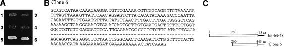
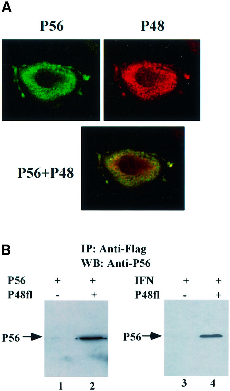
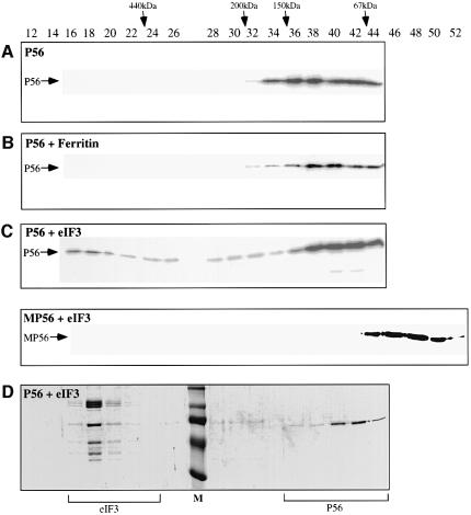
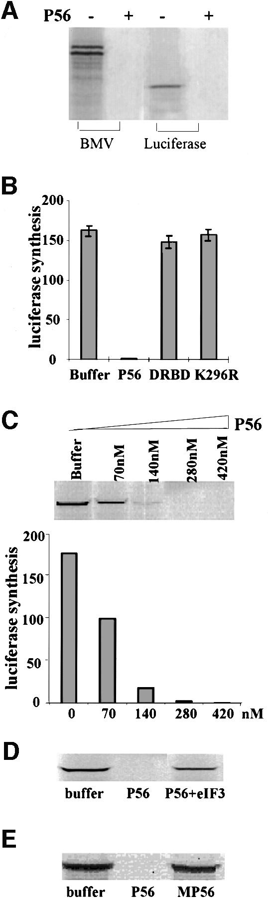
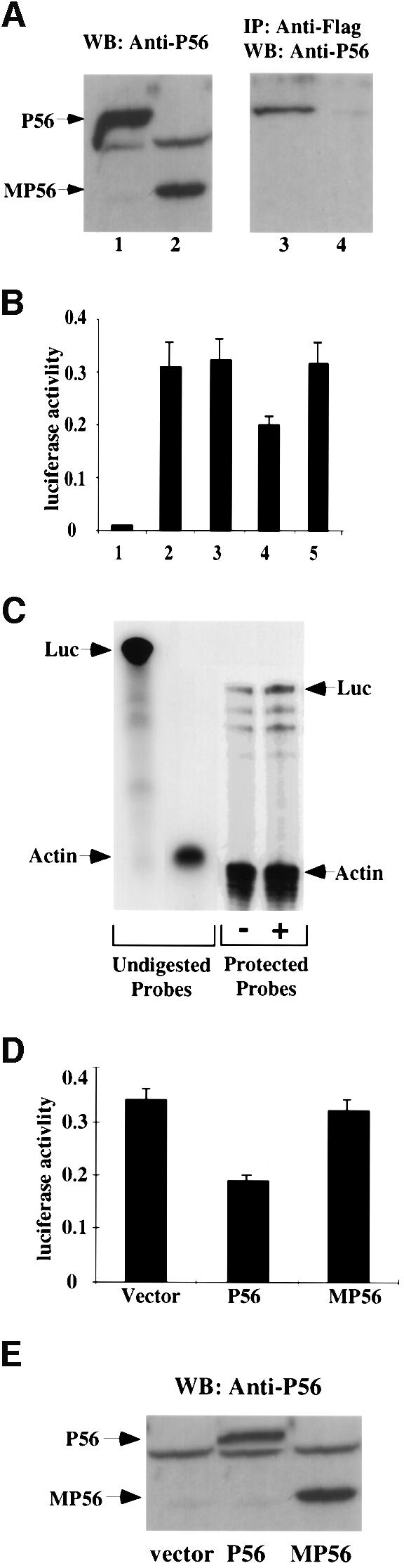
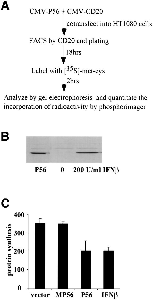
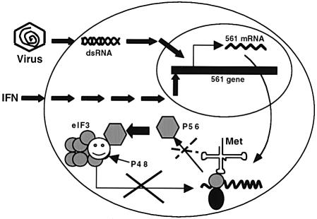
We report a new pathway of translation regulation that may operate in interferon-treated or virus-infected mammalian cells. This pathway is activated by P56, a protein whose synthesis is strongly induced by interferons or double-stranded RNA. Using a yeast two-hybrid screen, we identified the P48 subunit of the mammalian translation initiation factor eIF-3 as a protein that interacts with P56. The P56-P48 interaction was confirmed in human cells by co-immunoprecipitation assays and confocal microscopy. Gel filtration assays revealed that P56 binds to the large eIF-3 complex that contains P48. Purified recombinant P56 inhibited in vitro translation of reporter mRNAs in a dose-dependent fashion, and that inhibition was reversed by the addition of purified eIF-3. In vivo, expression of transfected P56 or induction of the endogenous P56 by interferon caused an inhibition of overall cellular protein synthesis and the synthesis of a transfected reporter protein. As expected, a P56 mutant that does not interact with P48 and eIF-3 failed to inhibit protein synthesis in vitro and in vivo.


Figures

None
Fig. 1. The interaction of clone 6 and P56 in yeast. (A) Yeast strain Y190 was co-transfected with the following pairs of expression vectors and plated onto the selection medium without histidine: (1) BD–vector + AD–clone 6; (2) BD–P56 + AD–SV40 large T-antigen; (3) BD–P56 + AD–vector; (4) BD–P53 + AD–clone 6; (5) BD–P56 + AD–clone 6; and (6) BD–P53 + AD–SV40 large T-antigen. (B) Partial cDNA sequence of clone 6. (C) Maps of full-length Int-6/P48 and clone 6.
None
Fig. 2. The interaction of P56 and Int-6/P48 in human cells. (A) Co-localization of P56 and P48 in the cytoplasm. HT1080 cells were transfected with pCMV-P48Fl and, 16 h post-transfection, cells were treated with 1000 U/ml IFN-β for 16 h. A confocal immunofluorescence assay was performed using anti-P56 antibody and anti-Flag antibody. The subcellular locations of P56 (green) and P48 (red) and their co-localization (yellow) are shown. (B) The interaction between both exogenous and endogenous P56 with P48. In lanes 1 and 2, HT1080 cells on a 100 mm plate were transfected with 10 µg of pCMV-P56 alone (lane 1) or co-transfected with 8 µg of pCMV-P56 and 8 µg of pCMV-P48Fl (lane 2). In lanes 3 and 4, cells were transfected with 8 µg of vector alone (lane 3) or pCMV-P48Fl (lane 4) and, 24 h post-transfection, cells were treated with 1000 U/ml IFN-β to induce endogenous P56. After 24 h, cell extracts were made and immunoprecipitation was performed with Flag antibody-conjugated Sepharose beads followed by western blotting with P56 antibody.
None
Fig. 3. The interaction between P56 and eIF-3 in vitro. Binding of P56 to eIF-3 was monitored by gel filtration chromatography. (A) Recombinant purified P56 protein (65 µg); (B) recombinant purified P56 protein (65 µg; mol. wt 56 kDa) and ferritin (261 µg; mol. wt 440 kDa) mixture (ferritin:P56 molar ratio = 1:2); and (C) recombinant purified P56 protein (65 µg; mol. wt 56 kDa) in the upper panel or recombinant purified MP56 protein (92 µg; mol. wt 40 kDa) in the lower panel was mixed with purified rabbit eIF-3 (350 µg; mol. wt 600 kDa) at a 1:2 molar ratio of eIF-3:P56 and a 1:4 molar ratio of eIF-3:MP56. (D) The same as the upper panel of (C). The different mixtures of proteins were analyzed by gel filtration chromatography on a Superdex 200 packed XK 16/70 column. In (A–C), 25 µl of each even fraction were used for western blotting analysis with P56 antibody. In (D), 500 µl of each even fraction were acetone precipitated and the precipitates were subjected to gel electrophoresis on a 10% SDS–polyacrylamide gel. The gel was run and stained with Coomassie Blue. Fraction numbers are indicated on the top. The positions of the different molecular weight markers are noted by arrows on the top. The positions of P56 detected by western blotting in (A–C) are indicated by arrows on the left. The fractions that contain eIF-3 or P56 are indicated at the bottom. M: pre-stained molecular weight marker.
None
Fig. 4. Inhibition of translation by P56 and rescuing the P56-mediated translation inhibition by eIF-3 in vitro. (A) Luciferase or BMV mRNA was translated in an in vitro translation system with or without 280 nM purified recombinant P56 protein. Synthesized BMV or luciferase was monitored by gel electrophoresis followed by fluorography. (B) Luciferase mRNA was translated in the presence of 280 nM recombinant purified P56, DRBD or K296R protein, or an equivalent volume of the dialysis buffer. The amounts of luciferase synthesized were quantified by phosphorimager analysis after gel electrophoresis. The averages of results from three experiments are shown. (C) Luciferase mRNA was translated in the presence of 70, 140, 280 or 420 nM recombinant purified P56 protein or an equivalent volume of the dialysis buffer. The synthesized luciferase was analyzed by gel electrophoresis and quantified by phosphorimager. (D) Purified rabbit eIF-3 (500 nM) and/or P56 (280 nM) was added to the translation system as indicated. Luciferase synthesis was analyzed by gel electrophoresis. (E) P56 (280 nM) or MP56 (280 nM) was added to the translation system as indicated and luciferase synthesis was analyzed by gel electrophoresis.
None
Fig. 5. Inhibition of synthesis of the luciferase reporter gene by P56 in vivo. (A) Interaction of P48/Int-6 with P56 but not MP56. pCMV-P56 (lanes 1 and 3) or pCMV-MP56 (lanes 2 and 4) was co-transfected with pCMV-P48Fl into cells. At 48 h post-transfection, cells were harvested and whole-cell extracts were prepared. A 50 µg aliquot of total cell protein was subjected to gel electrophoresis followed by western blotting with P56 antibody (lanes 1 and 2). A 1 mg aliquot of cell protein was subjected to immunoprecipitation with anti-Flag-conjugated Sepharose beads followed by western blot analysis with P56 antibody (lanes 3 and 4). (B) Cells were co-transfected with E-selectin-Luc and pCMV-P56 (bar 4), pCMV-MP56 (bar 5), pCMV-DRBP76 (bar 3) or the empty expression vector (bars 1 and 2). After 48 h, cells were treated with TNF-α (bars 2–5) for 4 h. Cell extracts were made and luciferase activity was measured. The averages of results from three experiments are shown. (C) Cells were co-transfected with E-selectin-Luc and pCMV-P56 (+) or vector (–). At 48 h post-transfection, cells were treated with TNF-α for 4 h. Cells were harvested and total RNA was isolated for RNase protection assay. A 40 µg aliquot of total RNA was hybridized with 32P-labeled Luc (370 bases) and γ-actin (140 bases) antisense RNA probes shown on the left as undigested probes. Following RNase digestion, the protected RNA probes were resolved in a 6% polyacrylamide, 8 M urea gel. Luciferase mRNA levels, shown on the right as protected probes, were quantified by phosphorimager and, after normalizing against the γ-actin mRNA levels, they were comparable in the two samples. (D) Cells were co-transfected with E-selectin-Luc and vector, pCMV-P56 or pCMV-MP56, as indicated. The experimental protocol was the same as in (B). (E) The same three cell extracts from (D) were western blotted with P56 antibody.
None
Fig. 6. Inhibition of overall protein synthesis by P56 in vivo. (A) The outline of the in vivo protein synthesis inhibition assay. (B and C) Cells were co-transfected with CMV-CD20 and pCMV-P56, pCMV-MP56 or vector. Transfected cells expressing CD20 were selected by FACS and plated. If IFN treatment was required, 4 h after plating, the sorted vector-transfected cells were treated with 200 U/ml IFN-β. (B) At 18 h after plating, cells were harvested and cell lysate was prepared. A 50 µg aliquot of total cell protein was subjected to gel electrophoresis followed by western blot analysis with P56 antibody. (C) At 18 h after plating, cells were labeled with [35S]methionine and cysteine labeling mix for 2 h. Cell extracts were made and equal amounts of protein were analyzed by gel electrophoresis. The level of radioactivity incorporated in all proteins was quantified by phosphorimager. The averages of results from three experiments are shown.
None
Fig. 7. Regulation of cellular protein synthesis by P56. Transcription of 561 mRNA is induced by IFN or virus/dsRNA using two different signaling pathways. P56, produced upon translation of 561 mRNA, binds to the P48 subunit of eIF-3 and blocks its function in peptide chain initiation. As a result, synthesis of cellular proteins, including that of P56 itself, is inhibited.

References

    1. Asano K., Merrick,W.C. and Hershey,J.W. (1997) The translation initiation factor eIF3-p48 subunit is encoded by int-6, a site of frequent integration by the mouse mammary tumor virus genome. J. Biol. Chem., 272, 23477–23480. - PubMed
    1. Askew G.R., Doetschman,T. and Lingrel,J.B. (1993) Site-directed point mutations in embryonic stem cells: a gene-targeting tag-and-exchange strategy. Mol. Cell. Biol., 13, 4115–4124. - PMC - PubMed
    1. Bandyopadhyay S.K., Kalvakolanu,D.V. and Sen,G.C. (1990) Gene induction by interferons: functional complementation between trans-acting factors induced by α interferon and γ interferon. Mol. Cell. Biol., 10, 5055–5063. - PMC - PubMed
    1. Bandyopadhyay S.K., Leonard,G.T.,Jr, Bandyopadhyay,T., Stark,G.R. and Sen,G.C. (1995) Transcriptional induction by double-stranded RNA is mediated by interferon-stimulated response elements without activation of interferon-stimulated gene factor 3. J. Biol. Chem., 270, 19624–19629. - PubMed
    1. Bluyssen H.A., Vlietstra,R.J., Faber,P.W., Smit,E.M., Hagemeijer,A. and Trapman,J. (1994) Structure, chromosome localization, and regulation of expression of the interferon-regulated mouse IFi54/IFi56 gene family. Genomics, 24, 137–148. - PubMed

Publication types

MeSH terms