

doi: 10.1093/emboj/19.24.6918.
DNA of Drosophila melanogaster contains 5-methylcytosine
Affiliations
- PMID: 11118227
- PMCID: PMC305887
- DOI: 10.1093/emboj/19.24.6918
Item in Clipboard
DNA of Drosophila melanogaster contains 5-methylcytosine
EMBO J.
.
Abstract
It is commonly accepted that the DNA of Drosophila melanogaster does not contain 5-methylcytosine, which is essential in the development of most eukaryotes. We have developed a new, highly specific and sensitive assay to detect the presence of 5-methylcytosine in genomic DNA. The DNA is degraded to nucleosides, 5-methylcytosine purified by HPLC and, for detection by 1D- and 2D-TLC, radiolabeled using deoxynucleoside kinase and [gamma-(32)P]ATP. Using this assay, we show here that 5-methylcytosine occurs in the DNA of D. melanogaster at a level of approximately 1 in 1000-2000 cytosine residues in adult flies. DNA methylation is detectable in all stages of D.melanogaster development.
Figures
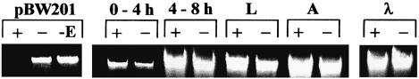
Fig. 1. McrBC cleavage of DNA isolated from D.melanogaster (+, with 1 mM GTP; –, in the absence of GTP). As a control, the cleavage of pBW201 plasmid DNA and λ DNA is shown. pBW201 is methylated at C CGG sequences at the first cytosine (+, with 1 mM GTP; –, in the absence of GTP; –E, without McrBC). λ DNA (MBI Fermentas) is not methylated at cytosine residues.
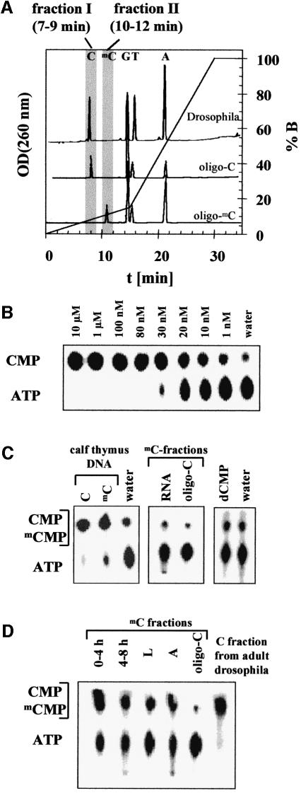
Fig. 2. (A) OD260 nm profiles of the HPLC separation of mononucleosides obtained from an enzymatic hydrolysis of DNA from adult D.melanogaster and two oligonucleotides used as control (oligo-C, which does not contain deoxymethylcytidine, and oligo-mC, which does not contain deoxycytidine). Two fractions were collected to quantify the deoxycytidine and deoxymethylcytidine content of the samples (C-fraction, 7–9 min; mC-fraction, 10–12 min). (B) Sensitivity of the enzymatic detection of deoxynucleosides using dNK. Different concentrations of deoxycytidine (Sigma) were incubated with dNK and analyzed by 1D-TLC as described in Materials and methods. The assay can detect 1 nM deoxycytidine in a 20 µl reaction volume corresponding to 20 fmol. (C) Control reactions of the HPLC/dNK assay. In the left panel, a 1D-TLC analysis of dNK-treated C- and mC-fractions of calf thymus DNA (Sigma) is shown. Methylcytidine is clearly detected in this reaction. In addition, 1D-TLC analyses of a dNK reaction using the mC-fraction of identically treated samples of yeast RNA (Boehringer Mannheim) and of dNK reactions with 200 nM dCMP are displayed, confirming that ribonucleosides and deoxyribonucleotides are not detected in the HPLC/dNK assay. (D) Detection of methylcytosine in DNA isolated from D.melanogaster at different stages of development (after 0–4 and 4–8 h of develop ment; L, from larvae; A, from adult flies). Shown are 1D-TLCs of dNK-treated mC-fractions. As a control, analyses of an identically treated sample of oligo-C that does not contain methylcytosine are shown. For comparison, the result obtained with C-fraction, which contains the cytidine, from adult flies is displayed.
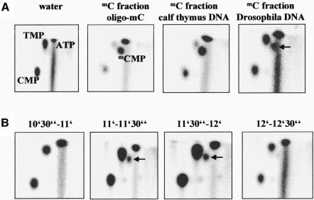
Fig. 3. (A) 2D-TLC analyses of mC-fractions of DNA from calf thymus, D.melanogaster and oligo-mC labeled with dNK. As a control, a 2D-TLC analysis of a reaction in which dNK is incubated with water is displayed. The relative positions of all four major nucleotides as well as of ATP were identified by 2D-TLCs with standard compounds. The mCMP spot in DNA isolated from D.melanogaster is marked with an arrow. (B) 2D-TLC analysis of the elution profile of methylcytidine from the HPLC column. In this experiment, DNA from adult flies was hydrolyzed and subjected to HPLC. Twenty fractions were collected between 5 and 15 min. In this figure, a 2D-TLC analysis of the fractions collected between 10.5–11, 11–11.5, 11.5–12 and 12–12.5 min is displayed, showing that only the fractions collected between 11 and 12 min contain methylcytidine. The mCMP spot in DNA isolated from D.melanogaster is marked with an arrow.
References
-
- Achwal C.W., Iyer,C.A. and Chandra,H.S. (1983) Immunochemical evidence for the presence of 5mC, 6mA and 7mG in human, Drosophila and mealybug DNA. FEBS Lett., 158, 353–358. - PubMed
-
- Adams R.L., McKay,E.L., Craig,L.M. and Burdon,R.H. (1979) Methylation of mosquito DNA. Biochim. Biophys. Acta, 563, 72–81. - PubMed
-
- Alberts B., Bray,D., Lewis,J., Raff,M., Roberts,K. and Watson,J.D. (1994) Molecular Biology of the Cell. Garland Publishing, Inc., New York, NY.
-
- Barbe J., Gibert,I. and Guerrero,R. (1986) 5-azacytidine: survival and induction of the SOS response in Escherichia coli K-12. Mutat. Res., 166, 9–16. - PubMed
Publication types
MeSH terms
Substances
LinkOut - more resources
Full Text Sources
Other Literature Sources
Molecular Biology Databases
