. 2000 Dec 15;529 Pt 3(Pt 3):661-8.
doi: 10.1111/j.1469-7793.2000.00661.x.

The effect of acidosis on systolic Ca2+ and sarcoplasmic reticulum calcium content in isolated rat ventricular myocytes

Affiliations

The effect of acidosis on systolic Ca2+ and sarcoplasmic reticulum calcium content in isolated rat ventricular myocytes

H S Choi et al. J Physiol. .

Abstract

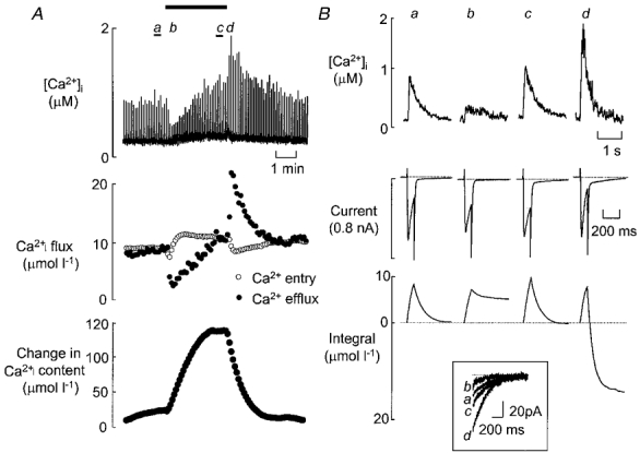
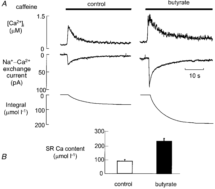
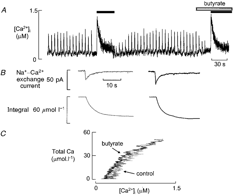
We have investigated the mechanisms responsible for the changes of systolic Ca2+ that occur in voltage-clamped rat ventricular myocytes during acidosis produced by application of the weak acid butyrate (30 mM). Intracellular pH regulation was inhibited with dimethylamiloride (bicarbonate-free solution). The application of butyrate produced an intracellular acidification of 0.33 pH units. This was accompanied by a decrease in systolic Ca2+ to about 50% of control. However, within 2 min, systolic Ca2+ returned to control levels. The decrease in systolic Ca2+ was accompanied by a decrease in the Na+-Ca2+ exchange current observed on repolarisation so that the calculated Ca2+ efflux on Na+-Ca2+ exchange was less than the entry on the L-type Ca2+ current. The magnitude of the Na+-Ca2+ exchange current recovered along with systolic Ca2+ until it equalled the Ca2+ entry on the L-type Ca2+ current. From the measurement of Ca2+ fluxes, it was calculated that, during acidosis, the cell gains 121.6+/-16.2 micromol l(-1) of Ca2+. This is equal to the measured increase of sarcoplasmic reticulum (SR) calcium content obtained by applying caffeine (20 mM) and integrating the resulting Na+-Ca2+ exchange current. We conclude that the recovery of the amplitude of the systolic Ca2+ transient is due to decreased SR calcium release, resulting in reduced Ca2+ efflux from the cell leading to increased SR calcium content.


Figures

Figure 1
Figure 1. The effect of butyrate (30 mM) on pHi (top) and [Ca2+]i (bottom) in representative rat ventricular myocytes
The cells were field stimulated at 0.2 Hz and exposed to butyrate for 2 min as shown above the traces. Different cells were used for the pH and [Ca2+]i measurements. In this (and all other) figures dimethylamiloride (20 μM) was present throughout to inhibit Na+-H+ exchange.
Figure 2
Figure 2. The effect of butyrate on transmembrane Ca2+ fluxes
A, time course. The top trace shows measurements of [Ca2+]i. The middle trace shows the calculated Ca2+ movements per pulse calculated from the L-type Ca2+ current (○) and Na+-Ca2+ exchange (•). The bottom trace shows the calculated change of cell calcium content obtained by summing the fluxes. Pulses 100 ms in duration were applied from -40 to 0 mV at 0.2 Hz. Butyrate (30 mM) was applied for the time shown by the horizontal bar. B, specimen traces obtained at the times indicated on A. From top to bottom, traces show: [Ca2+]i, membrane current, integrated calcium movement. An upward deflection on the integral trace represents Ca2+ entry and a downward one, Ca2+ exit. The inset at the bottom shows the current on repolarisation.
Figure 3
Figure 3. The effect of butyrate on sarcoplasmic reticulum calcium content
A, original data. Traces show, from top to bottom: [Ca2+]i, Na+-Ca2+ exchange current, and the integral of the Na+-Ca2+ exchange current. Caffeine was applied as shown by the horizontal bars. For the right-hand record butyrate (30 mM) was applied 2 min before the record began. B, mean data (6 cells) showing the effects of butyrate on SR calcium content.
Figure 4
Figure 4. Comparison of the effects of butyrate on systolic and caffeine-evoked Ca2+ transients
A, time course of effects on [Ca2+]i. The cell was stimulated with 100 ms duration depolarising pulses from -40 to 0 mV applied at 0.2 Hz. Stimulation was discontinued while caffeine was applied (black bar). Butyrate (30 mM) was applied for 20 s (open bar) before and during the second caffeine response. B, Na+-Ca2+ exchange current (top) and integral (bottom) produced by caffeine in control (left) and butyrate (right). C, Ca2+ buffering curves obtained in control and butyrate.

References

    1. Allen DG, Orchard CH. The effects of changes of pH on intracellular calcium transients in mammalian cardiac muscle. The Journal of Physiology. 1983;335:555–567. - PMC - PubMed
    1. Balnave CD, VaughanJones RD. Effect of intracellular pH on spontaneous Ca2+ spark frequency in rat ventricular myocytes. The Journal of Physiology. 2000;528:25–37. - PMC - PubMed
    1. Bountra C, VaughanJones RD. Effect of intracellular and extracellular pH on contraction in isolated mammalian cardiac tissue. The Journal of Physiology. 1989;418:163–187. - PMC - PubMed
    1. Buckler KJ, VaughanJones RD. Application of a new pH-sensitive fluoroprobe (carboxy-SNARF-1) for intracellular pH measurement in small, isolated cells. Pflügers Archiv. 1990;417:234–239. - PubMed
    1. Choi HS, Trafford AW, Eisner DA. Measurement of calcium entry and exit in quiescent rat ventricular myocytes. Pflügers Archiv. 2000;440:600–608. - PubMed

Publication types

MeSH terms

LinkOut - more resources