. 2001 Jan;69(1):194-203.
doi: 10.1128/IAI.69.1.194-203.2001.

Susceptibility to secondary Francisella tularensis live vaccine strain infection in B-cell-deficient mice is associated with neutrophilia but not with defects in specific T-cell-mediated immunity

Affiliations

Susceptibility to secondary Francisella tularensis live vaccine strain infection in B-cell-deficient mice is associated with neutrophilia but not with defects in specific T-cell-mediated immunity

C M Bosio et al. Infect Immun. 2001 Jan.

Abstract

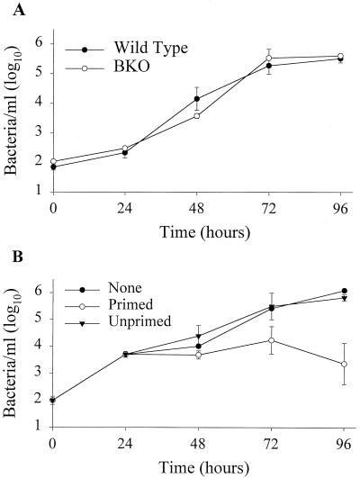
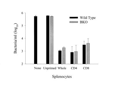
Previous studies have demonstrated a role for B cells, not associated with antibody production, in protection against lethal secondary infection of mice with Francisella tularensis live vaccine strain (LVS). However, the mechanism by which B cells contribute to this protection is not known. To study the specific role of B cells during secondary LVS infection, we developed an in vitro culture system that mimics many of the same characteristics of in vivo infection. Using this culture system, we showed that B cells do not directly control LVS infection but that control of LVS growth is mediated primarily by LVS-primed T cells. Importantly, B cells were not required for the generation of effective memory T cells since LVS-primed, B-cell-deficient (BKO) mice generated CD4(+) and CD8(+) T cells that controlled LVS infection similarly to LVS-primed CD4(+) and CD8(+) T cells from wild-type mice. The control of LVS growth appeared to depend primarily on gamma interferon and nitric oxide and was similar in wild-type and BKO mice. Rather, the inability of BKO mice to survive secondary LVS infection was associated with marked neutrophil influx into the spleen very early after challenge. The neutrophilia was directly associated with B cells, since BKO mice reconstituted with naive B cells prior to a secondary challenge with LVS had decreased bacterial loads and neutrophils in the spleen and survived.


Figures

FIG. 1
FIG. 1
Growth and control of LVS in BMMφ. (A) Growth of LVS in wild-type and BKO BMMφ. BMMφ from wild-type and BKO mice were infected with LVS at an MOI of 1:10 (bacterium-to-macrophage ratio). At the specified time points after infection, BMMφ were washed, lysed, and plated. Data points show the mean numbers ± the standard error of the mean (SEM) of viable bacteria (triplicate samples) recovered from wild-type (filled circles) and BKO (open circles) BMMφ at the indicated time points. (B) Control of LVS growth by LVS-primed splenocytes. Uninfected mice (Unprimed) or mice primed intradermally with LVS 4 weeks prior to the onset of the experiment (Primed) served as the source of LVS-primed splenocytes. Immediately following LVS infection of BMMφ, 5 × 10⁶ splenocytes of each population were added to designated wells. At the indicated time points, cultures were assessed for intracellular bacteria. Data points show the mean numbers ± the SEM of viable bacteria (triplicate samples) recovered from macrophages alone (None, filled circles), cultures containing primed splenocytes (open circles), or cultures containing unprimed splenocytes (filled triangles) at the indicated time points; the SEM was too small to be visualized for some of the data points on this graph. The data in panel A are representative of three experiments similar in design, and those in panel B are representative of five experiments similar in design.
FIG. 2
FIG. 2
Determination of specificity, concentration, and time of splenocyte addition to cultures for optimal control of LVS growth. In each graph, “None” represents LVS bacterial growth in BMMφ alone. (A) BMMφ were infected with LVS. Immediately or 24 h after infection, primed (black bars) or unprimed (gray bars) splenocytes (5 × 10⁶/well) were added to cultures. Seventy-two hours after infection, cultures were assessed for intracellular bacteria as described in the legend to Fig. 1. Error bars represent the SEM. (B) BMMφ were infected with LVS (gray bars) or L. monocytogenes (black bars); BMMφ were infected with L. monocytogenes at an MOI of 1:1,000 (bacterium-to-macrophage ratio). Twenty-four hours after infection of BMMφ splenocytes of each population (5 × 10⁶/well) were added to designated wells. Seventy-two hours after addition of splenocytes, cultures were assessed for intracellular bacteria as described in the legend to Fig. 1. (C) BMMφ were infected with LVS. Twenty-four hours after infection, the indicated concentrations of primed (black bars) or unprimed (gray bars) splenocytes were added to cultures. Seventy-two hours after infection, cultures were assessed for intracellular bacteria. These data are representative of two experiments similar in design.
FIG. 3
FIG. 3
Control of LVS growth by lymphocyte subpopulations. Uninfected mice served as a source of unprimed splenocytes. Mice primed intradermally with LVS 4 weeks prior to the onset of the experiment (Primed) served as the source of LVS-primed splenocytes. T and B lymphocytes were enriched from spleens of primed mice as described in Materials and Methods; efficacy of enrichment was determined by flow cytometry. Immediately following infection of BMMφ, unprimed splenocytes (5 × 10⁶/well), primed (Whole) splenocytes (5 × 10⁶/well), or primed T cells or B cells (2.3 × 10⁶/well) were added. Seventy-two hours after infection, cultures were assessed for intracellular bacteria. These results are representative of three experiments similar in design.
FIG. 4
FIG. 4
Growth and control of LVS in BMMφ by wild-type and BKO splenocytes. BMMφ from wild-type (A) or BKO (B) mice were infected with LVS. Immediately following infection, splenocytes from wild-type (5 × 10⁶/well) or BKO (2.5 × 10⁶/well) mice primed intradermally with LVS 4 weeks prior to the onset of the experiment were added to cultures. At the indicated time points after infection, cultures were assessed for intracellular bacteria. These results are representative of three experiments similar in design.
FIG. 5
FIG. 5
Control of LVS growth by T-lymphocyte subpopulations. Uninfected mice served as a source of unprimed splenocytes. Wild-type (black bars) and BKO (gray bars) mice primed intradermally with LVS 4 weeks prior to the onset of the experiment served as the sources of LVS-primed splenocytes. CD4+ and CD8+ T lymphocytes were enriched from the spleens of primed mice as described in Materials and Methods; efficacy of enrichment was determined by flow cytometry. Immediately following infection of BMMφ, the following were added to cultures: 5 × 10⁶ unprimed splenocytes/well; 5 × 10⁶ wild-type or 2.5 × 10⁶ BKO (Whole) primed splenocytes/well; and 1.3 × 10⁶ wild-type CD4+, 1 × 10⁶ wild-type CD8+, 1.1 × 10⁶ BKO CD4+, or 8.8 × 10⁵ BKO CD8+ T cells/well. Seventy-two hours after infection, cultures were assessed for intracellular bacteria. These results are representative of three experiments similar in design.
FIG. 6
FIG. 6
Secretion of cytokines and NO into culture supernatants following LVS infection. Splenocytes from either unprimed wild-type or BKO mice (gray bars) or mice primed with 8 × 10⁴ LVS 4 weeks prior to the onset of the experiment (black bars) were added to cultures of LVS-infected BMMφ. Culture supernatants (triplicate samples) were tested for IFN-γ (A), IL-12 (B), or NO (C) 72 h after addition of splenocytes. “None” represents LVS-infected BMMφ alone. ND, not detected. Each bar represents the mean ± the SEM cytokine or NO concentration of a group. These results are representative of three experiments similar in design.
FIG. 7
FIG. 7
Control of LVS in BMMφ by wild-type and BKO mouse splenocytes at various time points after priming. BMMφ were infected with LVS. Immediately following infection, splenocytes from wild-type or BKO mice primed intradermally with LVS 1, 2, 3, and 4 weeks prior to the onset of the experiment were added to cultures in cDMEM (A) or cDMEM containing anti-IFN-γ antibodies (10 μg/ml) (B) at 5 × 10⁶ or 2.5 × 10⁶ splenocytes/well, respectively. Seventy-two hours after infection, cultures were assessed for intracellular bacteria. The data shown are the mean numbers of viable bacteria ± the SEM recovered from macrophages alone (hatched bar) and cultures containing wild-type (black bars) or BKO (gray bars) splenocytes at the indicated time points. These results are representative of three experiments similar in design.
FIG. 8
FIG. 8
Secretion of cytokines and NO into culture supernatants following LVS infection. Splenocytes from wild-type (unfilled bars) or BKO (diagonal bars) mice primed intradermally with LVS 1, 2, 3, and 4 weeks prior to the onset of the experiment were added to cultures of LVS-infected BMMφ at 5 × 10⁶ or 2.5 × 10⁶ splenocytes/well, respectively. Culture supernatants (triplicate samples) were tested for IFN-γ (A), IL-12 (B), or NO (C) 72 h after addition of splenocytes. “None” represents LVS-infected BMMφ alone (black bars). ND, not detected. Each bar represents the mean concentration of cytokine or NO ± the SEM in a group. These results are representative of three experiments similar in design.
FIG. 9
FIG. 9
Splenocyte populations in wild-type and BKO mice 1, 2, 3, and 4 weeks following priming with LVS. Splenocytes from mice (three per group) were harvested, adhered to slides by cytocentrifugation, and stained for examination of cell morphology using a modified Wright-Giemsa stain. Cells categorized as lymphocytes, macrophages, and neutrophils are represented. Splenocytes from uninfected (Unprimed) mice are also shown. BKO mice had a significantly greater numbers of neutrophils 1, 2, and 3 weeks after priming compared to wild-type mice (P < 0.05). Wild-type mice had significantly more lymphocytes at each time point before and after priming compared to BKO mice (all P < 0.05). These results are representative of two experiments similar in design.
FIG. 10
FIG. 10
Growth of LVS and analysis of splenocyte populations in wild-type C57BL/6J mice; BKO mice, and reconstituted BKO mice following i.p. infection. Wild-type mice, BKO mice, and BKO mice that received 2.3 × 10⁷ naive B cells prior to infection were studied. All mice were primed intradermally with 8 × 10⁴ LVS bacteria 4 weeks prior to i.p. infection. (A) Splenocyte populations in wild-type, BKO, and reconstituted BKO mice following i.p. challenge with 5 × 10⁵ LVS bacteria. Splenocytes from mice were harvested, adhered to slides by cytocentrifugation, and stained for examination of cell morphology using a modified Wright-Giemsa stain. Cells categorized as lymphocytes, macrophages, and neutrophils are represented. BKO mice had significantly greater numbers of neutrophils 1 and 2 days after infection compared to wild-type and reconstituted BKO mice (P < 0.05). These results are representative of two experiments similar in design. (B) Growth of LVS in organs following i.p. challenge. The data shown are the mean numbers ± the SEM of viable bacteria recovered from the spleens, lungs, and livers at the indicated time points. ⧫, statistically significant difference (P < 0.05) compared to wild-type mice; ∗, all remaining mice dead by day 3 after infection. Although not represented here, BKO mice had statistically significantly greater numbers of bacteria in the spleens and lungs compared to reconstituted mice 1 day after infection. Both wild-type and reconstituted mice survived for more than 90 days following infection.

References

    1. Anthony L S D, Burke R D, Nano F E. Growth of Francisella spp. in rodent macrophages. Infect Immun. 1991;59:3291–3296. - PMC - PubMed
    1. Anthony L S D, Ghadirian E, Nestel F P, Kongshavn P A L. The requirement for gamma interferon in resistance of mice to experimental tularemia. Microb Pathog. 1989;7:421–428. - PubMed
    1. Anthony L S D, Kongshavn P A L. Experimental murine tularemia caused by Francisella tularensis, live vaccine strain: a model of acquired cellular resistance. Microb Pathog. 1987;2:3–14. - PubMed
    1. Conlan J W, North R J. Neutrophil-mediated dissolution of infected host cells as a defense strategy against a facultative intracellular bacterium. J Exp Med. 1991;174:741–744. - PMC - PubMed
    1. Conlan J W, North R J. Early pathogenesis of infection in the liver with the facultative intracellular bacteria Listeria monocytogenes, Francisella tularensis, and Salmonella typhimurium involves lysis of infected hepatocytes by leukocytes. Infect Immun. 1992;60:5164–5171. - PMC - PubMed

Publication types

LinkOut - more resources