. 2001 Jan;75(1):311-22.
doi: 10.1128/JVI.75.1.311-322.2001.

Kinetics of hepadnavirus loss from the liver during inhibition of viral DNA synthesis

Affiliations

Kinetics of hepadnavirus loss from the liver during inhibition of viral DNA synthesis

Y Zhu et al. J Virol. 2001 Jan.

Abstract

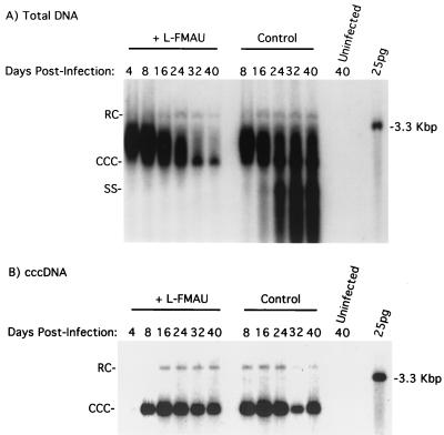
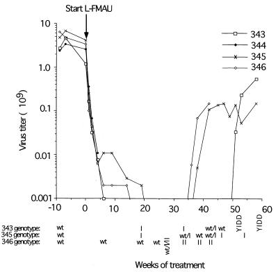
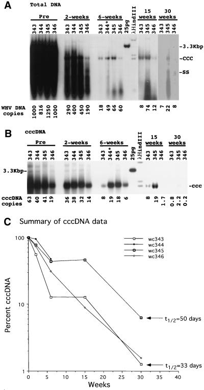
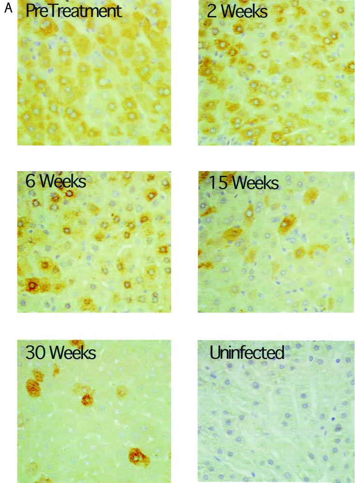
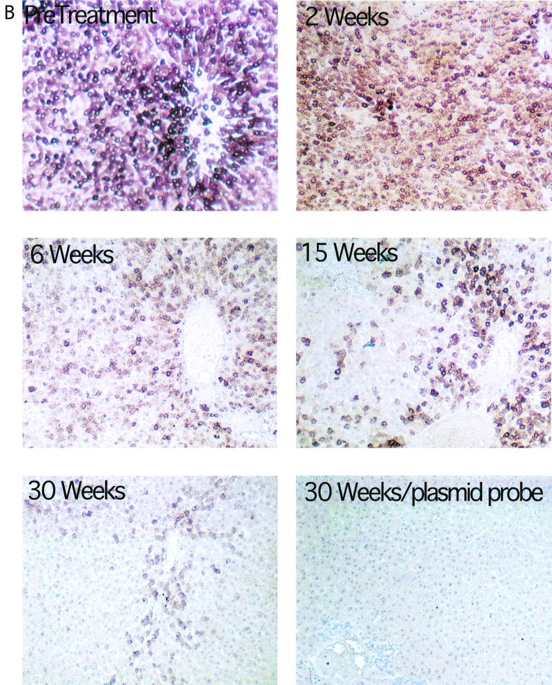
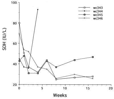
Hepadnaviruses replicate by reverse transcription, which takes place in the cytoplasm of the infected hepatocyte. Viral RNAs, including the pregenome, are transcribed from a covalently closed circular (ccc) viral DNA that is found in the nucleus. Inhibitors of the viral reverse transcriptase can block new DNA synthesis but have no direct effect on the up to 50 or more copies of cccDNA that maintain the infected state. Thus, during antiviral therapy, the rates of loss of cccDNA, infected hepatocytes (1 or more molecules of cccDNA), and replicating DNAs may be quite different. In the present study, we asked how these losses compared when woodchucks chronically infected with woodchuck hepatitis virus were treated with L-FMAU [1-(2-fluoro-5-methyl-beta-L-arabinofuranosyl) uracil], an inhibitor of viral DNA synthesis. Viremia was suppressed for at least 8 months, after which drug-resistant virus began replicating to high titers. In addition, replicating viral DNAs were virtually absent from the liver after 6 weeks of treatment. In contrast, cccDNA declined more slowly, consistent with a half-life of approximately 33 to 50 days. The loss of cccDNA was comparable to that expected from the estimated death rate of hepatocytes in these woodchucks, suggesting that death of infected cells was one of the major routes for elimination of cccDNA. However, the decline in the actual number of infected hepatocytes lagged behind the decline in cccDNA, so that the average cccDNA copy number in infected cells dropped during the early phase of therapy. This observation was consistent with the possibility that some fraction of cccDNA was distributed to daughter cells in those infected hepatocytes that passed through mitosis.


Figures

FIG. 1
FIG. 1
L-FMAU does not induce loss of cccDNA from primary cultures of WHV-infected woodchuck hepatocytes. Hepatocyte cultures were infected and treated with L-FMAU, beginning 4 days postinfection, as described in Materials and Methods. Total DNA (A) and cccDNA (B) were extracted from the cells at the indicated times and subjected to Southern blot analysis. Each lane contained one-quarter of the DNA extracted from a 6-cm-diameter tissue culture dish. RC, relaxed circular 3.3-kbp DNA.
FIG. 2
FIG. 2
Suppression and rebound of viremia during long-term treatment with L-FMAU. Virus titers during treatment with L-FMAU were quantified by Southern blot assays for virus particles in the serum, as described in Materials and Methods. A titer of 106 DNA equivalents per ml represents the lower limit of detection of our assays. Serum samples for genotype analysis were collected 9 weeks before and 6, 19, 24, 27, 33, 38, 42, 45, 51, 53, and 58 weeks after initiation of therapy. The changes in the sequences of the B and C domains of the viral DNA polymerase which define the mutant designations were determined by direct sequencing of PCR products and are illustrated in Fig. 5. wt, wild type.
FIG. 3
FIG. 3
Loss of replicating and cccDNA forms of viral DNA during antiviral therapy. Viral DNA was extracted from liver biopsy specimens for analysis of total and cccDNA forms of viral DNA by Southern blot analysis on 1.5% agarose gels, as described in Materials and Methods. Each lane contained either 10 μg of total DNA (A) or a cccDNA-enriched fraction recovered from 106 liver cells (based on nuclear counts in the liver lysates) (B). The average copy numbers of viral DNA per hepatocyte, shown at the bottom of panels A and B, were calculated as equivalents of full-length, double-stranded viral DNA and assume that the liver comprises 70% hepatocytes. The genome of the woodchuck was assumed to weigh 5 pg per diploid cell. The percent loss of cccDNA during treatment is summarized in panel C. The arrows indicate the losses of cccDNA after 30 weeks that would occur via a first-order decay with the indicated half-lives. Woodchuck 344 (wc344) died after 4 weeks of therapy.
FIG. 4
FIG. 4
Progressive loss of hepatocytes with detectable levels of WHV core antigen and nucleic acids during antiviral therapy. All liver biopsy specimens were assayed for the fraction of hepatocytes with detectable levels of WHV core antigen and nucleic acids. The results of these analyses for serial liver biopsies from woodchuck 346 are illustrated. Core antigen was detected by an immunoperoxidase assay (A), and viral nucleic acids were detected by in situ hybridization (B). The percentage of positive hepatocytes detected with each assay is summarized in Table 1. Magnification: (A) ×200; (B) ×100. Uninfected liver tissue is shown as a negative control in panel A. In panel B, tissue hybridized with a plasmid-specific probe (no WHV insert) served as the negative control.
FIG. 4
FIG. 4
Progressive loss of hepatocytes with detectable levels of WHV core antigen and nucleic acids during antiviral therapy. All liver biopsy specimens were assayed for the fraction of hepatocytes with detectable levels of WHV core antigen and nucleic acids. The results of these analyses for serial liver biopsies from woodchuck 346 are illustrated. Core antigen was detected by an immunoperoxidase assay (A), and viral nucleic acids were detected by in situ hybridization (B). The percentage of positive hepatocytes detected with each assay is summarized in Table 1. Magnification: (A) ×200; (B) ×100. Uninfected liver tissue is shown as a negative control in panel A. In panel B, tissue hybridized with a plasmid-specific probe (no WHV insert) served as the negative control.
FIG. 5
FIG. 5
WHV active-site mutations.
FIG. 6
FIG. 6
Evidence that mutations that confer lamivudine-resistant DNA synthesis on a laboratory strain of WHV also confer resistance to L-FMAU. Resistance of viral DNA synthesis to L-FMAU was assayed in transfected HepG2 cells, as described in Materials and Methods. The predicted amino acid sequences of the DNA polymerase B and C domains of the type I, II, and III mutations are illustrated in Fig. 5. Evidence that these mutations confer lamivudine resistance has already been published (45).
FIG. 7
FIG. 7
Evidence that in vivo infections that are resistant to lamivudine may also be resistant to L-FMAU. WHV-infected woodchucks in which virus titers had rebounded during a previous treatment with lamivudine received a 7-week treatment with L-FMAU (A and B); control animals received the Dyets formula as a placebo (C and D). Virus titers and genotypes in the interval between the end of the lamivudine therapy and the beginning of L-FMAU or placebo administration are shown in panels A and C. In panels A and C, the first serum sample was collected on the day that lamivudine treatment was discontinued. Sequence determinations from direct sequencing of PCR products were validated by cloning and sequencing of PCR products from the zero time points for woodchucks (wc) 326, 331, 336, 342, 335, and 338, as well as the 2-week time point from woodchuck 335. The variants are listed according to their relative abundance in the PCR products, the first being the most abundant. The predicted amino acid sequences of the B and C domains of the DNA polymerase of the various mutants are summarized in Fig. 5. wt, wild type.
FIG. 8
FIG. 8
L-FMAU therapy did not induce a rise in serum SDH levels. Serum samples collected during the first 16 weeks of therapy were assayed for SDH, expressed in international units (IU) per liter (20). The level of SDH in chronically infected woodchucks is generally below 80 IU. No significant elevation was observed except in one woodchuck (wc344) at the time of death, apparently from sepsis unrelated to drug administration.

References

    1. Aguesse-Germon S, Liu S H, Chevallier M, Pichoud C, Jamard C, Borel C, Chu C K, Trepo C, Cheng Y C, Zoulim F. Inhibitory effect of 2′-fluoro-5-methyl-beta-l-arabinofuranosyl-uracil on duck hepatitis B virus replication. Antimicrob Agents Chemother. 1998;42:369–376. - PMC - PubMed
    1. Aldrich C E, Coates L, Wu T T, Newbold J, Tennant B C, Summers J, Seeger C, Mason W S. In vitro infection of woodchuck hepatocytes with woodchuck hepatitis virus and ground squirrel hepatitis virus. Virology. 1989;172:247–252. - PubMed
    1. Allen M I, Deslauriers M, Andrews C W, Tipples G A, Walters K A, Tyrrell D L, Brown N, Condreay L D. Identification and characterization of mutations in hepatitis B virus resistant to lamivudine. Lamivudine Clinical Investigation Group. Hepatology. 1998;27:1670–1677. - PubMed
    1. Barker L F, Chisari F V, McGrath P P, Dalgard D W, Kirschstein R L, Almeida J D, Edgington T S, Sharp D G, Peterson M R. Transmission of type B viral hepatitis to chimpanzees. J Infect Dis. 1973;127:648–652. - PubMed
    1. Bartholomeusz A, Groenen L C, Locarnini S A. Clinical experience with famciclovir against hepatitis B virus and development of resistance. Intervirology. 1997;40:337–342. - PubMed

Publication types

LinkOut - more resources