. 2001 Jan;75(1):362-74.
doi: 10.1128/JVI.75.1.362-374.2001.

Functional analysis of the simian immunodeficiency virus Vpx protein: identification of packaging determinants and a novel nuclear targeting domain

Affiliations

Functional analysis of the simian immunodeficiency virus Vpx protein: identification of packaging determinants and a novel nuclear targeting domain

S Mahalingam et al. J Virol. 2001 Jan.

Abstract

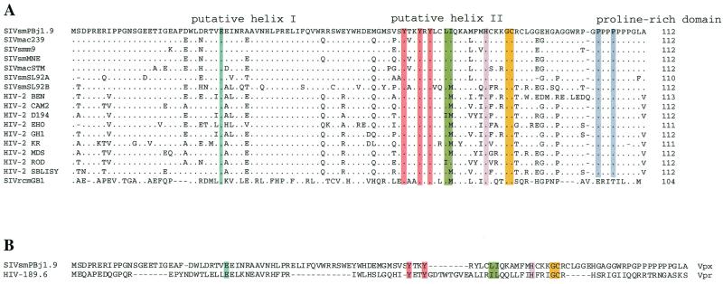
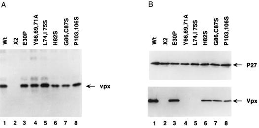
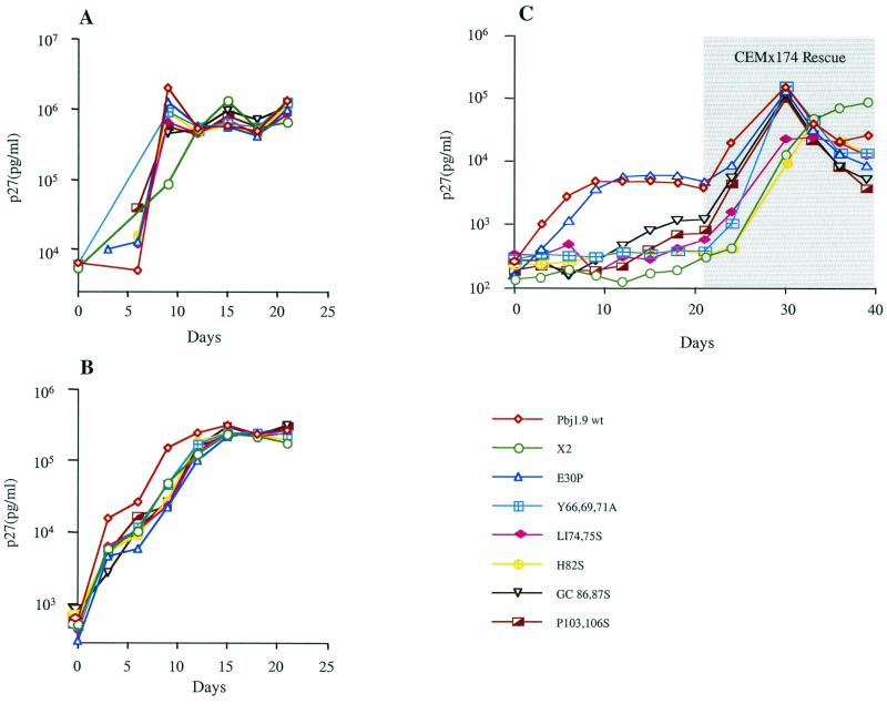
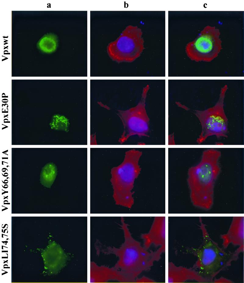
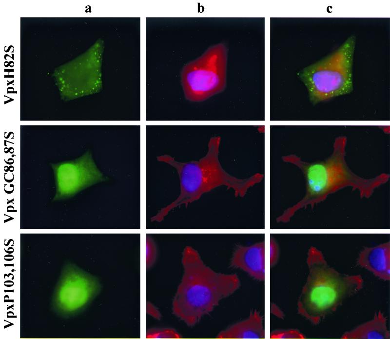
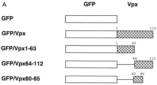
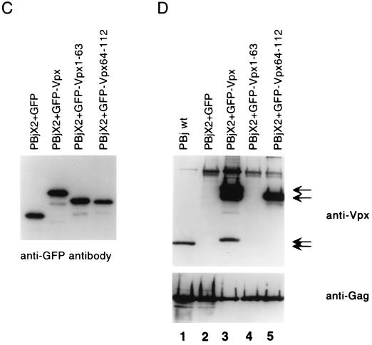
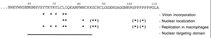
The vpx gene products of human immunodeficiency virus type 2 (HIV-2) and of the closely related simian immunodeficiency viruses from sooty mangabeys (SIVsm) and macaques (SIVmac) comprise a 112-amino-acid virion-associated protein that is critical for efficient virus replication in nondividing cells such as macrophages. When expressed in the absence of other viral proteins, Vpx localizes to the nuclear membrane as well as to the nucleus; however, in the context of virus replication Vpx is packaged into virions via interaction with the p6 domain of the Gag precursor polyprotein (p55(gag)). To identify the domains essential for virion incorporation and nuclear localization, site-directed mutations were introduced into the vpx gene of SIVsmPBj1.9 and functionally analyzed. Our results show that (i) mutation of two highly conserved L74 and I75 residues impaired both virion incorporation and nuclear localization of Vpx; (ii) substitution of conserved H82, G86, C87, P103, and P106 residues impaired Vpx nuclear localization but not virion incorporation; (iii) mutations of conserved Y66, Y69, and Y71 residues impaired virion incorporation but not the translocation of Vpx to the nucleus; and (iv) a mutation at E30 (predicted to disrupt an N-terminal alpha-helix) had no effect on either virion incorporation or nuclear localization of Vpx. Importantly, mutations in Vpx which impaired nuclear localization also reduced virus replication in macaque macrophages, suggesting an important role of the carboxyl terminus of Vpx in nuclear translocation of the viral preintegration complex. Analyzing this domain in greater detail, we identified a 26-amino-acid (aa 60 to 85) fragment that was sufficient to mediate the transport of a heterologous protein (green fluorescent protein [GFP]) to the nucleus. Taken together, these results indicate that virion incorporation and nuclear localization are encoded by two partially overlapping domains in the C-terminus of Vpx (aa 60 to 112). The identification of a novel 26-amino-acid nuclear targeting domain provides a new tool to investigate the nuclear import of the HIV-2/SIV preintegration complex.


Figures

FIG. 1
FIG. 1
(A) Alignment of deduced Vpx protein sequences from divergent HIV-2 and SIV isolates. Sequences are compared to the SIVsm(PBj1.9) Vpx protein sequence. Dots indicate amino acid sequence identity. Dashes represent gaps inserted to improve the alignment. The HIV-2/SIV Vpx sequences shown were obtained from the Los Alamos HIV sequence database (http://hiv-web.lanl.gov/), except for the deduced Vpx protein sequence of SIVrcm(GB1), which is unpublished. (B) Alignment of Vpx [SIVsm(PBj1.9)] and Vpr [HIV-1(89.6)] protein sequences. Groups of amino acid residues chosen for mutagenesis are highlighted in color.
FIG. 2
FIG. 2
Construction of SIVsm(PBj1.9) vpx mutant proviruses. The genetic organization of the SIVsm(PBj1.9) genome is shown at the top, with Vpx substitution mutations indicated at the bottom. None of the amino acid substitutions altered the coding sequences of the overlapping Vif open reading frames.
FIG. 3
FIG. 3
Expression and virion incorporation of mutant Vpx proteins. (A) vTF7-3 infected HeLa cells were transfected with Vpx expression plasmids. Transfected cells were labeled with 35S and the cell-associated Vpx proteins were immunoprecipitated with a Vpx-specific monoclonal antiserum. Lane 1, wild-type (Wt); lane 2, X2 (a control construct lacking a functional vpx open reading frame) (12); lanes 3 to 8, mutant constructs, as indicated above lanes. (B) Western blot analysis of SIVsm(PBj1.9) viral particles containing mutant Vpx proteins. 293T cells were transfected with vpx mutant PBj1.9 proviral clones. Virus particles were concentrated from culture supernatants by ultracentrifugation through a 20% sucrose cushion, solubilized in gel loading buffer, and analyzed for protein content by Western blot analysis using Gag (upper panel)- and Vpx (lower panel)-specific antibodies. Lane 1, wild type (Wt); lane 2, negative control (see legend for panel A); lanes 3 to 8, mutant constructs, as indicated above lanes.
FIG. 4
FIG. 4
Replication kinetics of wild-type and vpx mutant PBj1.9 proviruses. CEMx174 cells (A), macaque PBMCs (B), and terminally differentiated macaque macrophages (C) were infected with the indicated SIVsm(PBj1.9) virus constructs equilibrated by p27gag content (10 ng of p27gag per 106 cells). The isolation and infection of primary macaque PBMCs and macrophages are described in Materials and Methods. Virus replication was assessed by quantifying the amounts of p27gag antigen in culture supernatants at 3-day intervals postinfection. Twenty-one days after infection, adherent macrophages were cocultured for 24 h with 1 × 106 CEMx174 cells. Nonadherent cells were removed and analyzed at 3-day intervals for p27gag antigen production. wt, wild type; X2, negative control.
FIG. 5
FIG. 5
Subcellular localization of Vpx. vTF7-3 infected HeLa cells were transfected with wild-type and mutant Vpx expression plasmids. Twenty-four hours following transfection, the expressed Vpx proteins were detected by indirect immunofluorescence with an anti-Vpx monoclonal antibody (74) followed by an anti-mouse FITC-conjugated secondary antibody. (a) Localization of Vpx; (b) staining of cytoplasmic (red) and nuclear (blue) compartments with Texas red-phalloidin and DAPI, respectively; (c) superimposition of images shown in panels a and b for each row.
FIG. 5
FIG. 5
Subcellular localization of Vpx. vTF7-3 infected HeLa cells were transfected with wild-type and mutant Vpx expression plasmids. Twenty-four hours following transfection, the expressed Vpx proteins were detected by indirect immunofluorescence with an anti-Vpx monoclonal antibody (74) followed by an anti-mouse FITC-conjugated secondary antibody. (a) Localization of Vpx; (b) staining of cytoplasmic (red) and nuclear (blue) compartments with Texas red-phalloidin and DAPI, respectively; (c) superimposition of images shown in panels a and b for each row.
FIG. 6
FIG. 6
Construction, subcellular localization, expression and packaging of GFP-Vpx fusion proteins. (A) Schematic representation of GFP-Vpx fusion proteins. (B) Subcellular localization of GFP-Vpx fusion proteins in HeLa cells (see Results for details). Subpanels: a, the nuclear membrane was visualized by indirect immunofluorescence using a nucleoporin p62 specific polyclonal antibody, followed by a Texas red-conjugated goat anti-rabbit secondary antibody (red), and DAPI was used for nuclear staining (blue); b, the GFP signal was used to localize the GFP-Vpx fusion proteins; c, superimposition of images shown in subpanels a and b for each row. (C) Expression of GFP-Vpx fusion proteins. 293T cells were transfected with various GFP-Vpx expression plasmids. Lysates of transfected cells were prepared 48 h after transfection and analyzed for fusion protein content by Western blot analysis using an anti-GFP antibody. (D) Packaging of GFP-Vpx fusion proteins. Expression plasmids were cotransfected with the PBj1.9 Vpx proviral clone X2, and virus particles were concentrated from culture supernatants by ultracentrifugation through a 20% sucrose cushion, solubilized in gel loading buffer, and analyzed for protein content by Western blot analysis using Vpx (top)- and Gag (bottom)-specific monoclonal antibodies. Arrows indicate proteins with Vpx reactivity. Lane 1, wild-type Vpx protein (18 kDa); lane 2, negative (GFP) control; lane 3, full-length GFP-Vpx fusion protein (46 kDa) as well as two smaller proteins (38 kDa and 20 kDa, respectively) likely representing protease cleavage products; lane 4, lack of GFP-Vpx1-63 packaging; lane 5, GFP-Vpx64-112 fusion protein (38 kDa).
FIG. 6
FIG. 6
Construction, subcellular localization, expression and packaging of GFP-Vpx fusion proteins. (A) Schematic representation of GFP-Vpx fusion proteins. (B) Subcellular localization of GFP-Vpx fusion proteins in HeLa cells (see Results for details). Subpanels: a, the nuclear membrane was visualized by indirect immunofluorescence using a nucleoporin p62 specific polyclonal antibody, followed by a Texas red-conjugated goat anti-rabbit secondary antibody (red), and DAPI was used for nuclear staining (blue); b, the GFP signal was used to localize the GFP-Vpx fusion proteins; c, superimposition of images shown in subpanels a and b for each row. (C) Expression of GFP-Vpx fusion proteins. 293T cells were transfected with various GFP-Vpx expression plasmids. Lysates of transfected cells were prepared 48 h after transfection and analyzed for fusion protein content by Western blot analysis using an anti-GFP antibody. (D) Packaging of GFP-Vpx fusion proteins. Expression plasmids were cotransfected with the PBj1.9 Vpx proviral clone X2, and virus particles were concentrated from culture supernatants by ultracentrifugation through a 20% sucrose cushion, solubilized in gel loading buffer, and analyzed for protein content by Western blot analysis using Vpx (top)- and Gag (bottom)-specific monoclonal antibodies. Arrows indicate proteins with Vpx reactivity. Lane 1, wild-type Vpx protein (18 kDa); lane 2, negative (GFP) control; lane 3, full-length GFP-Vpx fusion protein (46 kDa) as well as two smaller proteins (38 kDa and 20 kDa, respectively) likely representing protease cleavage products; lane 4, lack of GFP-Vpx1-63 packaging; lane 5, GFP-Vpx64-112 fusion protein (38 kDa).
FIG. 7
FIG. 7
Mutational analysis of the C-terminal half of Vpx. The asterisks indicate amino acid residues that are absolutely essential for the functions indicated; the brackets denote residues that (when mutated) cause some functional impairment. The horizontal bar highlights the fragment of Vpx that is sufficient to mediate the transport of a heterologous protein (GFP) to the nucleus.

References

    1. Accola M A, Bukovsky A A, Jones M S, Göttlinger H G. A conserved dileucine-containing motif in p6gag governs the particle association of Vpx and Vpr of simian immunodeficiency viruses SIVmac and SIVagm. J Virol. 1999;73:9992–9999. - PMC - PubMed
    1. Bonifaci N, Moroianu J, Radu A, Blobel G. Karyopherin beta2 mediates nuclear import of a mRNA binding protein. Proc Natl Acad Sci USA. 1997;94:5055–5060. - PMC - PubMed
    1. Bresnahan P A, Yonemoto W, Greene W C. Cutting edge: SIV Nef protein utilizes both leucine- and tyrosine-based protein sorting pathways for down-regulation of CD4. J Immunol. 1999;163:2977–2981. - PubMed
    1. Bukrinsky M I, Haggerty S, Dempsey M P, Sharova N, Adzhubel A, Spitz L, Lewis P, Goldfarb D, Emerman M, Stevenson M. A nuclear localization signal within HIV-1 matrix protein that governs infection of non-dividing cells. Nature. 1993;365:666–669. - PMC - PubMed
    1. Chou P Y, Fasman G D. Prediction of the secondary structure of proteins from their amino acid sequence. Adv Enzymol Relat Areas Mol Biol. 1978;47:45–48. - PubMed

Publication types

MeSH terms

Substances