. 2001 Jan;75(1):384-95.
doi: 10.1128/JVI.75.1.384-395.2001.

An Epstein-Barr virus protein interacts with Notch

Affiliations

An Epstein-Barr virus protein interacts with Notch

S Kusano et al. J Virol. 2001 Jan.

Abstract

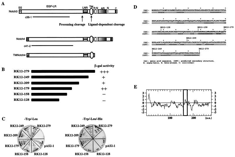
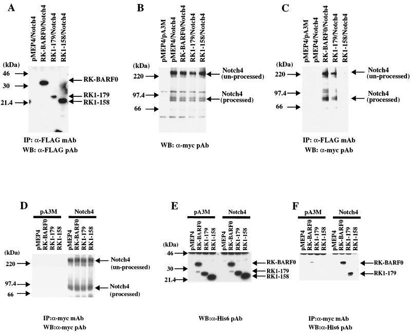
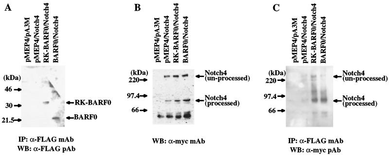
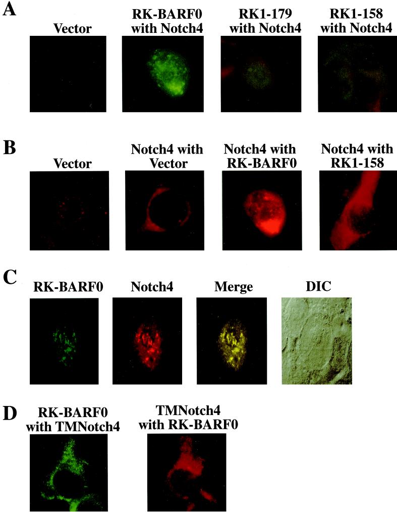
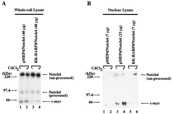
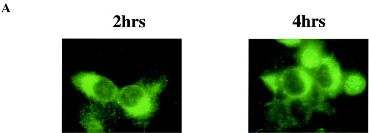
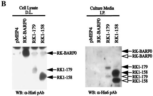
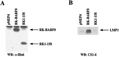
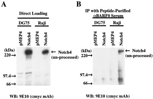
The Epstein-Barr virus (EBV) BamHI A mRNAs were originally identified in cDNA libraries from nasopharyngeal carcinoma, where they are expressed at high levels. The RNAs are differentially spliced to form several open reading frames and also contain the BARF0 open reading frame at the 3' end. One cDNA, RK-BARF0, included a potential endoplasmic reticulum-targeting signal peptide sequence. The RK-BARF0 protein is shown here to interact with the Notch4 ligand binding domain, using yeast two-hybrid screening, coimmunoprecipitation, and confocal microscopy. This interaction induces translocation of a portion of the full-length unprocessed Notch4 to the nucleus by using the Notch nuclear localization signal. These effects of RK-BARF0 on Notch intracellular location indicate that EBV possibly modulates Notch signaling. Unprocessed Notch4 was also detected in immunoprecipitated complexes from EBV-infected cells by using a rabbit antiserum raised against a BARF0-specific peptide. This finding provides additional evidence for expression of RK-BARF0 and its interaction with Notch during EBV infection. In EBV-infected, EBNA2-negative cells, RK-BARF0 induced the expression of EBV latent membrane protein 1 (LMP1), and this induction was dependent on the RK-BARF0/Notch interaction domain. The activation of LMP1 expression by RK-BARF0 may be responsible for expression of LMP1 in EBV latent infections in the absence of EBNA2.


Figures

FIG. 1
FIG. 1
(A) Schematic representation of the Notch clones obtained from two-hybrid screening. Clones containing the EGF-like repeat regions of Notch3 (c36-1) and Notch4 (c41-2) interacted with RK-BARF0 by yeast two-hybrid screening. A schematic representation of the Notch4 mutant lacking the entire functional intracellular region (TMNotch4) that is used this study is shown. The sites of the proteolytic cleavage for processing and ligand-dependent cleavage are indicated. Structural motifs within the Notch proteins are indicated as follows: SS, signal sequence; EGF-LR, EGF-like repeats; LNR, Notch/lin-12 like repeats; TM, transmembrane domain; R, RAM domain; N, NLS; AR, cdc10/ankyrin repeats; P, PEST sequence. (B) Schematic representation of C-terminal deletion mutants of RK-BARF0 and β-gal activity in the yeast mating assay. The full-length (aa 12 to 279) and five C-terminal deletion (aa 12 to 249, 12 to 209, 12 to 179, 12 to 158, and 12 to 128) constructs of RK-BARF0 fused to GAL4BD were transfected in yeast Y190 cells and mated with yeast Y187 cells containing the Notch4 EGF-like repeats region/GAL4AD fusion construct. Mated yeast cells were plated on Trp Leu medium plates, and the relative β-gal activity was determined. The time taken for more than 80% of the colonies to become blue is shown: +++, <1 h; ++, 1 to 3 h; +, 3 to 6 h; −, no blue colonies is >8 h. (C) Growth patterns of mated yeast strains on His selective medium. Mated yeast cells were plated on Trp Leu medium plates and Trp Leu His medium plates and incubated at 30°C for 5 days. (D) The predicted secondary structure of RK-BARF0 was determined using a 3D-1D compatibility algorithm. Bold letters indicate the sequence of the interaction-domain of RK-BARF0 with Notch. (E) The predicted hydrophobicity of the RK-BARF0 protein was determined by Kyte-Doolittle analysis. Bold square indicates the hydrophobicity of the interaction domain of RK-BARF0 with Notch.
FIG. 2
FIG. 2
RK-BARF0 associates with processed and unprocessed forms of Notch4 in vivo. H1299 cells (5 × 10⁵) were transiently transfected with both FLAG/His6-tagged RK-BARF0 (aa 1 to 279), RK1–179, RK1–158, or pMEP4 and Notch4myc or pA3M. RK-BARF0, RK1–179, and RK1–158 were induced with 5 μM CdCl2 for 16 h, immunoprecipitated, and analyzed on immunoblots. (A) Immunoblot with an anti-FLAG antibody to identify RK-BARF0, RK1–179, and RK1–158 in the immunoprecipitated RK-BARF0 complexes. (B) Immunoblot analysis with anti-myc antibody to identify Notch4 expression in the transfected cells. (C) Identification of processed and unprocessed forms of Notch4 in the immunoprecipitated (IP) RK-BARF0 complexes using anti-myc epitope polyclonal antibodies. (D) Immunoblot with anti-myc antibody to identify Notch4 in the immunoprecipitated Notch4 complexes. (E) Immunoblot analysis with antihistidine antiserum to determine the expression of RK-BARF0, RK1–179, and RK1–158 in the transfected cells. (F) Detection of RK-BARF0 and RK1–179, using antihistidine antibody in the Notch4 complexes immunoprecipitated with anti-myc antibody.
FIG. 3
FIG. 3
BARF0 interacts with processed Notch. (A) Immunoblot of RK-BARF0 or BARF0 (aa 106 to 279) after induction and immunoprecipitation (IP) with an anti-FLAG polyclonal antibody. (B) Immunoblot analysis of whole cell lysates to identify Notch4 expression after transfection. (C) Identification of processed and unprocessed forms of Notch4 in the RK-BARF0 and BARF0 immunoprecipitated complexes. WB, Western blot.
FIG. 4
FIG. 4
RK-BARF0 Colocalizes with Notch4 in the nucleus. (A) Cell lines containing both Notch4-myc and FLAG/His6-tagged RK-BARF0, RK1–179, RK1–158 or pMEP4 expression plasmids were induced and stained using a monoclonal antibody to FLAG and FITC-conjugated anti-mouse immunoglobulin G (IgG) to identify RK-BARF0. (B) Cell lines containing Notch4-myc or both Notch4-myc and FLAG/His6-tagged RK-BARF0, RK1–158, or the pMEP4 vector were stained with anti-myc polyclonal antibody and LSRC-conjugated anti-rabbit IgG to identify Notch4. (C) A stable cell line containing Notch4-myc and FLAG/His6-tagged RK-BARF0 was stained with anti-FLAG M2 monoclonal antibody and anti-myc polyclonal antibody using FITC-conjugated anti-mouse IgG to detect RK-BARF0 and RISC-conjugated anti-rabbit IgG to detect Notch4. Confocal images were collected, and composite figures were created using Adobe Photoshop 5.0.2. The differential interference contrast microscopy image of the cell is presented. (D) A cell line containing the extracellular to transmembrane region of Notch4 (TMNotch4)-myc in pA3M and FLAG/His6-tagged RK-BARF0 was stained with a monoclonal FLAG and FITC-conjugated anti-mouse IgG to identify RK-BARF0 and anti-myc polyclonal antibody and LSRC-conjugated anti-rabbit IgG to identify TMNotch4.
FIG. 5
FIG. 5
RK-BARF0 induces the translocation of unprocessed Notch4 to the nucleus. Stable cell clones containing FLAG/His6 epitope-tagged RK-BARF0 or pMEP4 expression plasmids were transiently transfected with Notch4-myc and treated for 24 h with 8 μM CdCl2 or left untreated. Protein (40 μg) of whole-cell lysates (A) and 7 or 23 μg of nuclear lysates (B) were subjected to SDS-PAGE and analyzed for the presence of Notch4-myc by using anti-myc (9E10) antibody.
FIG. 6
FIG. 6
RK-BARF0 is expressed in the ER and can be secreted. (A) Stable H1299 cell lines containing FLAG/His6-tagged RK-BARF0 were induced by 5 μM CdCl2 to express RK-BARF0, and cells were stained 2 and 4 h postinduction with anti-FLAG antibody and FITC-conjugated anti-mouse antibody to determine RK-BARF0 localization. (B) Culture medium (8 ml/5 × 106 cells) of induced P3HR1 cells containing FLAG/His6-tagged RK-BARF0, RK1–179, RK1–158, or pMEP4 expression plasmids was incubated with anti-FLAG M2 beads. Immunoprecipitates (I.P.) from culture medium and 50 μg of protein of whole-cell lysates were subjected to SDS-PAGE. FLAG/His6-tagged RK-BARF0, RK1–179, or RK1–158 were detected with the anti-His- probe polyclonal antibody. Solid arrows indicate the protein bands which are identified in both whole-cell lysate and cell culture media; open arrows indicate the protein bands which are present only in the cell culture media. D.L., direct load; WB, Western blot.
FIG. 6
FIG. 6
RK-BARF0 is expressed in the ER and can be secreted. (A) Stable H1299 cell lines containing FLAG/His6-tagged RK-BARF0 were induced by 5 μM CdCl2 to express RK-BARF0, and cells were stained 2 and 4 h postinduction with anti-FLAG antibody and FITC-conjugated anti-mouse antibody to determine RK-BARF0 localization. (B) Culture medium (8 ml/5 × 106 cells) of induced P3HR1 cells containing FLAG/His6-tagged RK-BARF0, RK1–179, RK1–158, or pMEP4 expression plasmids was incubated with anti-FLAG M2 beads. Immunoprecipitates (I.P.) from culture medium and 50 μg of protein of whole-cell lysates were subjected to SDS-PAGE. FLAG/His6-tagged RK-BARF0, RK1–179, or RK1–158 were detected with the anti-His- probe polyclonal antibody. Solid arrows indicate the protein bands which are identified in both whole-cell lysate and cell culture media; open arrows indicate the protein bands which are present only in the cell culture media. D.L., direct load; WB, Western blot.
FIG. 7
FIG. 7
RK-BARF0 induces LMP1 expression in EBNA2-negative P3HR1 cells. Stable cell lines containing FLAG/His6-tagged RK-BARF0, RK1–158, or pMEP4 expression plasmids were treated for 48 h with 1.5 μM CdCl2. (A) Immunoblot analysis of 80 μg of protein of whole-cell lysates with an anti-His-probe to identify RK-BARF0 and RK1–158. (B) Immunoblot analysis of 60 μg of protein of whole-cell lysates by anti-LMP1 (CS1-4) to identify LMP1. WB, Western blot.
FIG. 8
FIG. 8
RK-BARF0 peptide rabbit antiserum precipitates transfected Notch4 protein from Raji cells but not from DG75. Stable cell lines containing myc-tagged Notch4 or pMEP4 expression plasmids were treated for 36 h with 5 μM CdCl2. (A) Protein (100 μg) from cell lysates was subjected to SDS-PAGE, and myc-tagged Notch4 was identified using the 9E10 monoclonal antibody. (B) Protein from cell lysates (1,250 μg) was precipitated using peptide affinity-purified rabbit antiserum against RK-BARF0 (10). Immunoprecipitates (IP) were subjected to SDS-PAGE, and myc-tagged Notch4 was identified using 9E10 monoclonal antibody. WB, Western blot.

References

    1. Aster J C, Robertson E S, Hasserjian R P, Turner J R, Kieff E, Sklar J. Oncogenic forms of NOTCH1 lacking either the primary binding site for RBP-Jkappa or nuclear localization sequences retain the ability to associate with RBP-Jkappa and activate transcription. J Biol Chem. 1997;272:11336–11343. - PubMed
    1. Blaumueller C M, Qi H, Zagouras P, Artavanis-Tsakonas S. Intracellular cleavage of Notch leads to a heterodimeric receptor on the plasma membrane. Cell. 1997;90:281–291. - PubMed
    1. Brooks L A, Lear A L, Young L S, Rickinson A B. Transcripts from the Epstein-Barr virus BamHI A fragment are detectable in all three forms of virus latency. J Virol. 1993;67:3182–3190. - PMC - PubMed
    1. Busson P, McCoy R, Sadler R, Gilligan K, Tursz T, Raab-Traub N. Consistent transcription of the Epstein-Barr virus LMP2 gene in nasopharyngeal carcinoma. J Virol. 1992;66:3257–3262. - PMC - PubMed
    1. Cohen J I, Wang F, Mannick J, Kieff E. Epstein-Barr virus nuclear protein 2 is a key determinant of lymphocyte transformation. Proc Natl Acad Sci USA. 1989;86:9558–9562. - PMC - PubMed

Publication types

Substances

LinkOut - more resources