. 2001 Jan;75(1):408-19.
doi: 10.1128/JVI.75.1.408-419.2001.

Interaction of the influenza virus nucleoprotein with the cellular CRM1-mediated nuclear export pathway

Affiliations

Interaction of the influenza virus nucleoprotein with the cellular CRM1-mediated nuclear export pathway

D Elton et al. J Virol. 2001 Jan.

Abstract

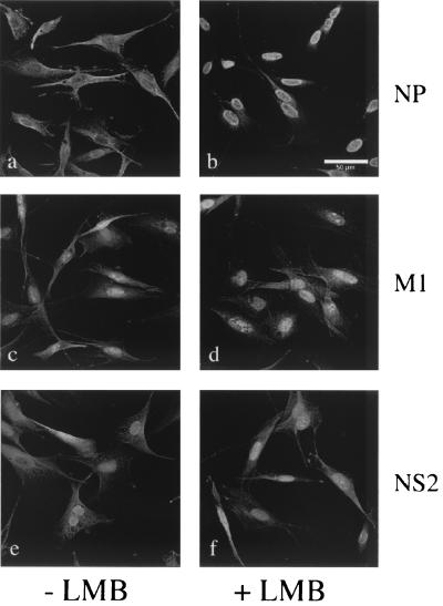
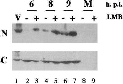
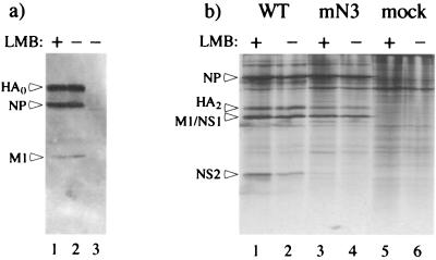
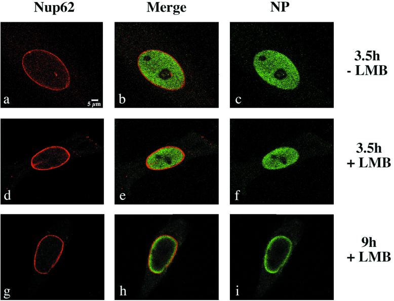
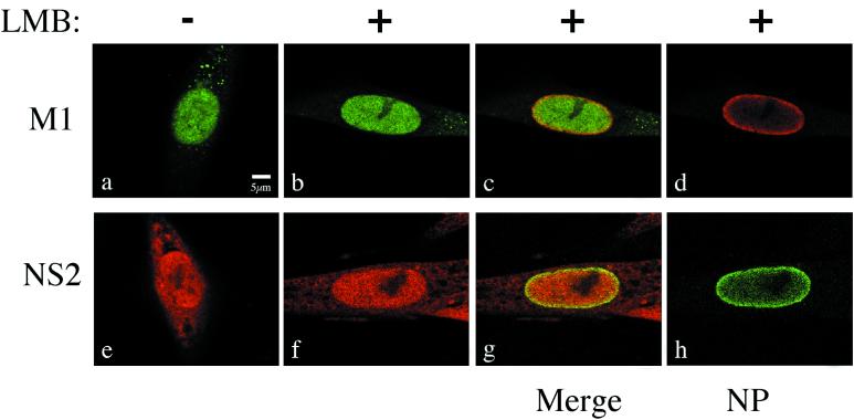
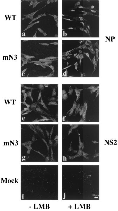
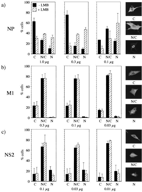
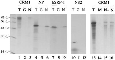
Influenza virus transcription occurs in the nuclei of infected cells, where the viral genomic RNAs are complexed with a nucleoprotein (NP) to form ribonucleoprotein (RNP) structures. Prior to assembly into progeny virions, these RNPs exit the nucleus and accumulate in the cytoplasm. The mechanisms responsible for RNP export are only partially understood but have been proposed to involve the viral M1 and NS2 polypeptides. We found that the drug leptomycin B (LMB), which specifically inactivates the cellular CRM1 polypeptide, caused nuclear retention of NP in virus-infected cells, indicating a role for the CRM1 nuclear export pathway in RNP egress. However, no alteration was seen in the cellular distribution of M1 or NS2, even in the case of a mutant virus which synthesizes greatly reduced amounts of NS2. Furthermore, NP was distributed throughout the nuclei of infected cells at early times postinfection but, when retained in the nucleus at late times by LMB treatment, was redistributed to the periphery of the nucleoplasm. No such change was seen in the nuclear distribution of M1 or NS2 after drug treatment. Similar to the behavior of NP, M1 and NS2 in infected cells, LMB treatment of cells expressing each polypeptide in isolation caused nuclear retention of NP but not M1 or NS2. Conversely, overexpression of CRM1 caused increased cytoplasmic accumulation of NP but had little effect on M1 or NS2 distribution. Consistent with this, NP bound CRM1 in vitro. Overall, these data raise the possibility that RNP export is mediated by a direct interaction between NP and the cellular CRM1 export pathway.


Figures

FIG. 1
FIG. 1
Effect of LMB treatment on the intracellular localization of influenza virus polypeptides. BHK cells were infected with 10 PFU of influenza A/PR/8/34 virus per cell; incubated from 1 hpi in the absence (a, c, and e) or presence (b, d, and f) of 11 nM LMB, and examined at 12 hpi by confocal microscopy after staining for NP (a and b), M1 (c and d), or NS2 (e and f). Images were generated using an extended-focus algorithm.
FIG. 2
FIG. 2
Effect of LMB on the distribution of viral RNPs between the nuclear and cytoplasmic cell fractions. BHK cells were infected with PR8 at an MOI of 10 PFU/cell, incubated from 1 hpi in the presence or absence of 11 nM LMB, and harvested at 6, 8, and 9 h postinfection. Cells were separated into nuclear (N) and cytosolic (C) fractions, and equivalent amounts were subjected to SDS-PAGE and Western blotting with anti-RNP serum. Samples of purified virus (lane v) were also analyzed in parallel to provide a marker for NP. M, mock infected.
FIG. 3
FIG. 3
Effect of LMB on influenza virus polypeptide synthesis. (a) BHK cells were infected with PR8 in the presence (lane 1) or absence (lane 2) of 11 nM LMB (added at 1 hpi), harvested at 13 hpi, and analyzed by SDS-PAGE and Western blotting using anti-PR8 serum. Mock-infected cell lysate is shown in lane 3. The positions of uncleaved HA, NP, and M1 are indicated by arrows. (b) CEF cells were infected at 34°C with WT FPV or the ts mutant mN3 in the presence (+) or absence (−) of LMB (added at 1 hpi) and then labeled with [35S]methionine for 30 min before harvesting at 8 hpi. Lysates were analyzed by SDS-PAGE and autoradiography. The positions of NP, HA2, M1/NS1, and NS2 are indicated.
FIG. 4
FIG. 4
Effect of LMB on the intranuclear distribution of NP. BHK cells were infected with PR8 in the absence or presence of 11 nM LMB (added at 1 hpi) and harvested at the indicated times postinfection. Cells were stained for NP (green) and Nup62 (red), and single optical planes of focus were examined by confocal microscopy. The green channels in panels h and i were captured at lower sensitivity to keep the maximum fluorescence intensity within the linear range of the detector.
FIG. 5
FIG. 5
Effect of LMB on the intranuclear distribution of M1 and NS2. BHK cells were infected with PR8 in the absence (−) or presence (+) of 11 nM LMB and harvested at 9 hpi. Cells were stained for either M1 (green) or NS2 (red) and costained for NP. Single optical planes of focus captured by confocal microscopy are shown. Panels b, c, and d and f, g, and h, respectively, are of the same cells.
FIG. 6
FIG. 6
Effect of LMB treatment on the intracellular localization of NP and NS2 in FPV-infected cells. CEF cells were infected (or mock infected) with either WT or mN3 FPV at 34°C, incubated from 1 hpi in the absence or presence of 11 nM LMB, and harvested at 8 hpi. Cells were stained for NP (a to d) or NS2 (e to j) and examined by confocal microscopy. Images were generated using an extended-focus algorithm.
FIG. 7
FIG. 7
Effect of LMB on the distribution of NP, NS2, and M1 in transfected cells. BHK cells were infected with vTF7 at an MOI of 10 PFU/cell and then transfected 2 h later with the indicated amounts of plasmid DNA encoding NP, NS2, or M1. At 3 h posttransfection, cells were overlaid with fresh medium containing either 11 nM LMB or no drug and harvested 2 h later. Cells were analyzed by immunofluorescence assay using antibodies for NP, M1, or NS2 and scored as follows for the localization pattern of the influenza virus protein: N, predominantly nuclear; N/C distributed throughout the cell; C, predominantly cytoplasmic. Examples of the staining patterns are shown on the right. The number of cells showing each distribution pattern was expressed as a percentage of the total cell count. The average and range of two independent experiments are shown.
FIG. 8
FIG. 8
In vitro interaction between NP and CRM1. Radiolabeled in vitro-translated CRM1, NP, hSRP-1, and NS2 were analyzed by SDS-PAGE and autoradiography before (T) or after binding to GST (G), MBP (M), or GST-NP (N in lanes 3, 6, 9, and 12) or MBP-NP (N in lanes 15 and 16) immobilized on agarose beads. LMB (11 nM) was included in the reaction mixture run in lane 15 (N+). The values on the left are molecular mass markers (kilodaltons).

References

    1. Askjaer P, Heick Jensen T, Nilsson J, Engelmeier L, Kjems J. The specificity of the CRM1-Rev nuclear export signal interaction is mediated by RanGTP. J Biol Chem. 1998;273:33414–33422. - PubMed
    1. Boyle S M, Ruvolo V, Gupta A K, Swaminathan S. Association with the cellular export receptor CRM1 mediates function and intracellular localization of Epstein-Barr virus SM protein, a regulator of gene expression. J Virol. 1999;73:6872–6881. - PMC - PubMed
    1. Briedis D J, Conti G, Munn E A, Mahy B W. Migration of influenza virus-specific polypeptides from cytoplasm to nucleus of infected cells. Virology. 1981;111:154–164. - PubMed
    1. Bui M, Wills E G, Helenius A, Whittaker G R. Role of the influenza virus M1 protein in nuclear export of viral ribonucleoproteins. J Virol. 2000;74:1781–1786. - PMC - PubMed
    1. Bullido R, Gomez-Puertas P, Albo C, Portela A. Several protein regions contribute to determine the nuclear and cytoplasmic localization of the influenza A virus nucleopotein. J Gen Virol. 2000;81:135–142. - PubMed

Publication types

LinkOut - more resources