. 2000 Dec;106(12):1481-8.
doi: 10.1172/JCI11176.

TNF-alpha induces osteoclastogenesis by direct stimulation of macrophages exposed to permissive levels of RANK ligand

Affiliations

TNF-alpha induces osteoclastogenesis by direct stimulation of macrophages exposed to permissive levels of RANK ligand

J Lam et al. J Clin Invest. 2000 Dec.

Abstract

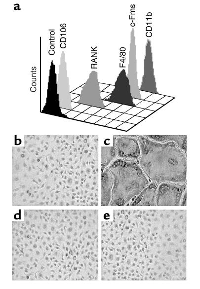
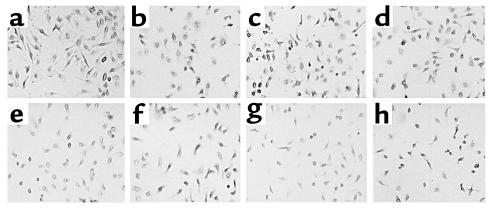
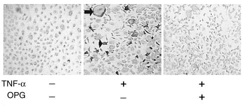
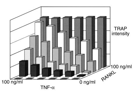
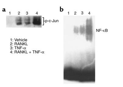
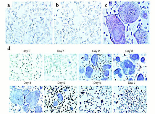
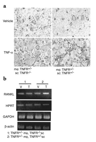
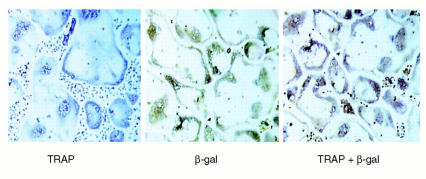
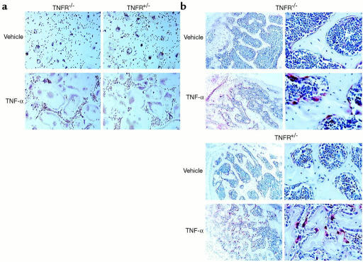
While TNF-alpha is pivotal to the pathogenesis of inflammatory osteolysis, the means by which it recruits osteoclasts and promotes bone destruction are unknown. We find that a pure population of murine osteoclast precursors fails to undergo osteoclastogenesis when treated with TNF-alpha alone. In contrast, the cytokine dramatically stimulates differentiation in macrophages primed by less than one percent of the amount of RANKL (ligand for the receptor activator of NF-kappaB) required to induce osteoclast formation. Mirroring their synergistic effects on osteoclast differentiation, TNF-alpha and RANKL markedly potentiate NF-kappaB and stress-activated protein kinase/c-Jun NH(2)-terminal kinase activity, two signaling pathways essential for osteoclastogenesis. In vivo administration of TNF-alpha prompts robust osteoclast formation in chimeric animals in which ss-galactosidase positive, TNF-responsive macrophages develop within a TNF-nonresponsive stromal environment. Thus, while TNF-alpha alone does not induce osteoclastogenesis, it does so both in vitro and in vivo by directly targeting macrophages within a stromal environment that expresses permissive levels of RANKL. Given the minuscule amount of RANKL sufficient to synergize with TNF-alpha to promote osteoclastogenesis, TNF-alpha appears to be a more convenient target in arresting inflammatory osteolysis.


Figures

Figure 1
Figure 1
Purified myeloid cells express macrophage lineage cell surface markers and differentiate into osteoclasts when cultured with RANKL. (a) Marrow cells were isolated by immunoselection and analyzed by flow cytometry. All purified cells are negative for CD106, a stromal cell marker, and positive for the macrophage lineage markers F4/80, c-Fms, and CD11b. Furthermore, the purified cells express RANK, a cell-surface receptor which is necessary for osteoclast differentiation. Cells were cultured with (b) 10 ng/ml M-CSF alone, or in combination with: (c) 100 ng/ml RANKL; (d) 500 ng/ml OPG; and (e) 100 ng/ml RANKL and 500 ng/ml OPG. Cells treated with RANKL differentiate into a confluent layer of multinucleated osteoclasts (panel c), a process completely inhibited by concomitant treatment with OPG (panel e).
Figure 2
Figure 2
Purified myeloid cells fail to differentiate into osteoclasts in response to TNF-α. Cells, as characterized in Figure 1, were cultured with 10 ng/ml M-CSF plus (a) 1 ng/ml, (b) 2 ng/ml, (c) 5 ng/ml, (d) 10 ng/ml, (e) 20 ng/ml, (f) 40 ng/ml, (g) 80 ng/ml, and (h) 160 ng/ml TNF-α. Cells were stained for TRAP activity after 7 days. No multinucleated or TRAP-expressing cells are evident.
Figure 3
Figure 3
TNF-α–induced differentiation of macrophages into osteoclasts is RANKL-dependent. Whole marrow was cultured with 100 ng/ml M-CSF, according to the method of Kobayashi et al. (11) and Azuma et al. (12) in the continuous presence or absence of 500 ng/ml OPG. After 3 days, 100 ng/ml TNF-α was added to some cultures. TNF-α induces formation of multinuclear (arrow) and mononuclear (arrowhead) TRAP-expressing cells only in the absence of OPG.
Figure 4
Figure 4
TNF-α synergizes with RANKL to stimulate osteoclast differentiation. Pure populations of myeloid cells were treated with various combinations of TNF-α and RANKL. After 5 days the extent of osteoclastogenesis was expressed as a function of TRAP activity, as determined by a colorimetric assay. No osteoclast formation is induced in the absence of RANKL, while in its presence, TNF-α augments osteoclast differentiation in a dose-dependent manner. TNF-α potentiation of osteoclastogenesis is seen with all RANKL dosages beneath saturating levels.
Figure 5
Figure 5
TNF-α and RANKL synergistically activate SAPK/JNK and NF-κB. (a) Purified myeloid cells were treated with 1 ng/ml RANKL and/or 500 pg/ml TNF-α for 12 minutes. Following cell lysis, an in vitro kinase assay was performed for SAPK/JNK activity. (b) Purified myeloid cells were treated with 10 ng/ml RANKL and/or 1 ng/ml TNF-α for 15 minutes. Nuclear extracts were analyzed by electrophoretic mobility shift assay (EMSA), using an oligonucleotide containing the NF-κB binding site from the TNF-α promoter.
Figure 6
Figure 6
TNF-α potentiates subosteoclastogenic levels of RANKL in a time-dependent manner. Purified myeloid cells were treated with 10 ng/ml M-CSF plus (a) 1 ng/ml RANKL; (b) 1 ng/ml TNF-α; and (c) 1 ng/ml RANKL plus 1 ng/ml TNF-α. Cells were stained for TRAP activity 7 days after cytokine addition. No osteoclast formation is seen with the minimal dosage of RANKL or TNF-α alone, while exuberant osteoclastogenesis is evident when the cytokines act in concert. (d) Purified myeloid cells were treated with 1 ng/ml RANKL plus 10 ng/ml M-CSF. On successive days, 500 pg/ml TNF-α was added. Cells were stained for TRAP activity 5 days after addition of TNF-α. TNF-α–induced osteoclastogenesis maximizes when the cytokine is added 2–4 days following RANKL exposure.
Figure 7
Figure 7
TNF-α, in vitro, acts directly on osteoclast precursors to enhance osteoclastogenesis primed by basal levels of RANKL. (a) Purified TNFR-heterozygous macrophages (mφ) were cultured with purified TNFR-deficient or TNFR-heterozygous stromal cells (sc), in the presence of 10 ng/ml TNF-α or vehicle. Exuberant osteoclastogenesis occurs regardless of the ability of stromal cells to respond to TNF-α. (b) RANKL expression in these cultures was analyzed by RT-PCR. Expression of RANKL is elevated by administration of TNF-α (T) relative to vehicle (V) only in the cultures containing TNF-responsive stromal cells.
Figure 8
Figure 8
Rosa macrophages differentiate into β-galactosidase–positive, committed osteoclast precursors when transplanted into TNFR-heterozygous mice. Bone marrow transplantation was employed to create chimeric animals in which β-galactosidase–positive osteoclast precursors exist within the stromal environment of TNFR-heterozygous mice. Following engraftment, marrow cells were cultured for 7 days in osteoclastogenic conditions. Generated osteoclasts were stained for TRAP and β-galactosidase (β-gal) activity, alone and in combination. TRAP histochemical staining yields a purple reaction product, while β-galactosidase immunohistochemical staining yields a brown reaction product. Double staining for both TRAP and β-galactosidase yields combined reaction products.
Figure 9
Figure 9
TNF-α directly induces myeloid cells to differentiate into committed osteoclast precursors and mature osteoclasts in vivo, irrespective of the capacity of stromal cells to respond to the cytokine. Chimeric animals were created by bone marrow transplantation in which β-galactosidase–positive osteoclast precursors exist within the stromal environments of TNFR-deficient animals or their TNFR-heterozygous littermates. Following engraftment, the animals were depleted of T cells and administered 150 μg/kg/day TNF-α or vehicle for 5 days by subcutaneous injection. (a) Equal numbers of whole marrow cells were then cultured ex vivo for 7 days in osteoclastogenic conditions, and stained for TRAP and β-galactosidase activity in combination. (b) Decalcified sections of long bones, representative of data from six chimeric animals, were stained for TRAP activity (red reaction product) (left panels, ×100; right panels, ×250).

References

    1. Lannigan FJ, O’Higgins P, McPhie P. The cellular mechanism of ossicular erosion in chronic suppurative otitis media. J Laryngol Otol. 1993;107:12–16. - PubMed
    1. Baroukh B, Saffar JL. Identification of osteoclasts and their mononuclear precursors. A comparative histological and histochemical study in hamster periodontitis. J Periodontal Res. 1991;26:161–166. - PubMed
    1. Nair SP, et al. Bacterially induced bone destruction: mechanisms and misconceptions. Infect Immun. 1996;64:2371–2380. - PMC - PubMed
    1. Bom-van Noorloos AA, et al. Bacteroides gingivalis stimulates bone resorption via interleukin-1 production by mononuclear cells. J Clin Periodontol. 1990;17:409–413. - PubMed
    1. Birkedal-Hansen H. Role of cytokines and inflammatory mediators in tissue destruction. J Periodontal Res. 1993;28:500–510. - PubMed

Publication types

MeSH terms