. 2000 Dec;106(12):1489-99.
doi: 10.1172/JCI10498.

Clearance of Alzheimer's amyloid-ss(1-40) peptide from brain by LDL receptor-related protein-1 at the blood-brain barrier

Affiliations

Clearance of Alzheimer's amyloid-ss(1-40) peptide from brain by LDL receptor-related protein-1 at the blood-brain barrier

M Shibata et al. J Clin Invest. 2000 Dec.

Abstract

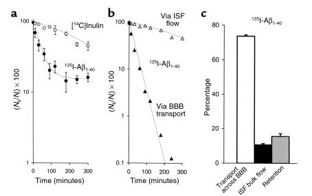
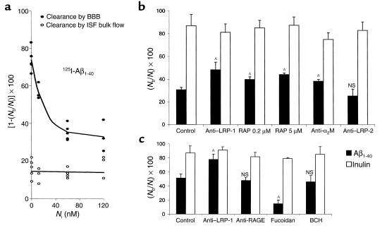
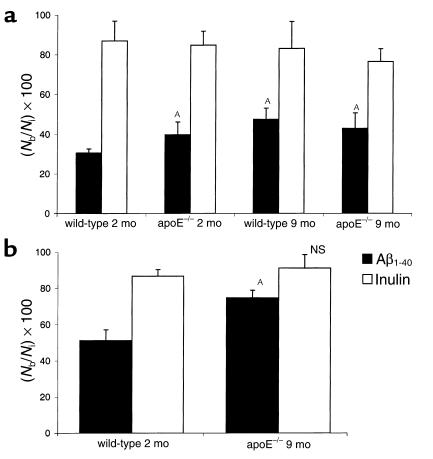
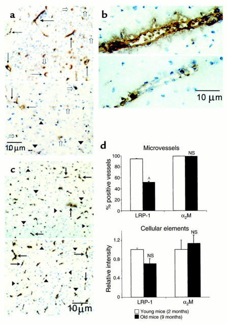
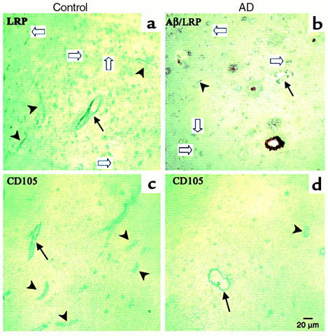
Elimination of amyloid-ss peptide (Ass) from the brain is poorly understood. After intracerebral microinjections in young mice, (125)I-Ass(1-40) was rapidly removed from the brain (t(1/2) </= 25 minutes), mainly by vascular transport across the blood-brain barrier (BBB). The efflux transport system for Ass(1-40) at the BBB was half saturated at 15.3 nM, and the maximal transport capacity was reached between 70 nM and 100 nM. Ass(1-40) clearance was substantially inhibited by the receptor-associated protein, and by antibodies against LDL receptor-related protein-1 (LRP-1) and alpha(2)-macroglobulin (alpha(2)M). As compared to adult wild-type mice, clearance was significantly reduced in young and old apolipoprotein E (apoE) knockout mice, and in old wild-type mice. There was no evidence that Ass was metabolized in brain interstitial fluid and degraded to smaller peptide fragments and amino acids before its transport across the BBB into the circulation. LRP-1, although abundant in brain microvessels in young mice, was downregulated in older animals, and this downregulation correlated with regional Ass accumulation in brains of Alzheimer's disease (AD) patients. We conclude that the BBB removes Ass from the brain largely via age-dependent, LRP-1-mediated transport that is influenced by alpha(2)M and/or apoE, and may be impaired in AD.


Figures

Figure 1
Figure 1
(a) Time-disappearance curves of [14C]inulin (open circles) and 125I-Aβ1-40 (60 nM; TCA-precipitable 125I radioactivity, filled circles) from the CNS after simultaneous microinjections of tracers into the caudate nucleus in mice. Each point represents the mean ± SD of three to seven animals. (b) Two components of 125I-Aβ1-40 efflux, vascular transport across the BBB (filled triangles) and transport via ISF bulk flow (open triangles), were computed with equations 3 and 4 using data from a. (c) Relative contributions to Aβ1-40 efflux by its transport across the BBB (open bar), diffusion via ISF bulk flow (filled bar), and retention (gray bar) in the brain were studied at 60 nM concentrations and calculated from the fractional coefficients given in Table 1.
Figure 2
Figure 2
Time-appearance curves of [14C]inulin (open circles) and 125I-Aβ1-40 (60 nM; TCA-precipitable 125I radioactivity, filled circles) in the CSF (a) and plasma (b) after simultaneous microinjections of tracers into the caudate nucleus in mice. Values are expressed as percentages of injected dose (%ID); each point is mean ± SD of three to seven animals.
Figure 3
Figure 3
(a) Brain TCA-precipitable (open bars) and non–TCA-precipitable 125I radioactivity (solid bars) after intracerebral microinjections of 125I-Aβ1-40 (60 nM) into the caudate nucleus in mice, expressed as a percentage of total 125I radioactivity in the brain; mean ± SD of three to five animals. (b) Left panel shows HPLC elution profile of brain tissue 60 minutes after intracerebral microinjection of 125I-Aβ1-40 (60 nM). Separation was performed for 30 mg of brain tissue on a reverse-phase HPLC column, using a 30-minute linear gradient of 25–83% acetonitrile in 0.1% TFA, pH 2. 125I-Aβ1-40 eluted at 52%, corresponding to the elution time of Aβ1-40 standard. Right panel shows SDS-PAGE analysis of brain tissue supernatant at 30 minutes (lane 1) and 60 minutes (lane 2) after intracerebral microinjection of 125I-Aβ1-40 (60 nM). The radioactivity in the brain eluted as a single peak on HPLC, with the same retention time as the Aβ1-40 standard (data not shown). Aliquots of lyophilized sample were subjected to 10% Tris-tricine SDS-PAGE, transferred to a nitrocellulose membrane, and exposed to x-ray film. (c) Plasma TCA-precipitable (open bars) and non–TCA-precipitable 125I radioactivity (filled bars) after intracerebral microinjections of 125I-Aβ1-40 (60 nM) into the caudate nucleus in mice, expressed as a percentage of total 125I radioactivity in plasma; mean ± SD of three to five animals.
Figure 4
Figure 4
(a) Concentration-dependent clearance of Aβ1-40 from mouse brain. Clearance via BBB transport (filled circles) is shown separately from clearance via ISF bulk flow (open circles). Clearance was determined 30 minutes after simultaneous microinjection of 125I-Aβ1-40 at increasing concentrations (0.05–120 nM) along with [14C]inulin into the caudate nucleus. (b) Effects of anti–LRP-1 Ab R777 (60 μg/ml), RAP (0.2 and 5 μM), anti-α2M Ab (20 μg/ml), and anti–LRP-2 Ab Rb6286 (60 μg/ml) on brain clearance of 125I-Aβ1-40 at 12 nM, determined 30 minutes after simultaneous microinjection of 125I-Aβ1-40 and [14C]inulin. (c) Effects of anti–LRP-1 Ab R777 (60 μg/ml), anti-RAGE Ab (60 μg/ml), fucoidin (100 μg/ml), and 2-amino-bicyclo[2.2.1]heptane-2-carboxylic acid (BCH; 10 mM) on brain clearance of 125I-Aβ1-40 at a higher load of 60 nM, determined 30 minutes after simultaneous microinjection of 125I-Aβ1-40 and [14C]inulin. Mean ± SD of three to four animals. AP < 0.05; NS, not significant.
Figure 5
Figure 5
(a) Effect of apoE genotype and age on brain clearance of 125I-Aβ1-40. Brain clearance of 125I-Aβ1-40 in 2-month-old and 9-month-old wild-type mice and apoE KO mice studied at the lower load of 12 nM 125I-Aβ1-40 (a) and a higher load of 60 nM (b). In all studies, 125I-Aβ1-40 and [14C]inulin were injected simultaneously, and clearance was determined after 30 minutes. Mean ± SD of three to four animals. AP < 0.05; NS, not significant compared with 2-month-old wild-type mice.
Figure 6
Figure 6
(a) LRP-1 immunoreactivity in brain microvessels of young (2-month-old; upper panel) and old (9-month-old; lower panel) wild-type mice. Many vessels in young mice stained positive for LRP-1, detected with anti–LRP-1 Ab R777 (5 μg/ml; arrows). There were relatively fewer positive vessels in old mice (arrows), and many weakly positive- or negative-staining vessels (arrowheads). There was no significant difference in the staining of parenchymal cellular elements (open arrows) between the young and old mice. Vessels in young mice stained strongly positive (b, upper panel) compared with the faint staining seen in old mice (b, lower panel). In contrast, there was no difference in staining for α2M in brain cells (arrowheads) or microvessels (arrows) between young (c, upper panel) and old (c, lower panel) mice. (d) Comparison of LRP-1 and α2M immunoreactivity in brain microvessels (upper panel) and parenchymal cellular elements (lower panel) in young and old wild-type mice. AP < 0.05; NS, not significant.
Figure 7
Figure 7
LRP-1 expression in human frontal cortex. Brain sections (Brodmann’s area 10) of controls (a and c) reveal well-defined staining of capillaries (arrowheads) and arterioles (arrows) by LRP-1, detected with anti–LRP-1 mAb 8G1 (5 μg/ml) (a) and CD105 (c). No Aβ staining was present in double-labeled or serially labeled sections (not shown). In contrast, double-labeled sections from AD patients show vessels and plaque cores stained positive with anti-Aβ1-40 (brown stain), reduced numbers and intensity of LRP-1 staining of vessels (b), and reduced numbers of CD105-labeled vessels (d).

References

    1. Wisniewski T, Ghiso J, Frangione B. Biology of Aβ amyloid in Alzheimer’s disease. Neurobiol Dis. 1997;4:311–328. - PubMed
    1. Selkoe DJ. Alzheimer’s disease: genotype, phenotype, and treatments. Science. 1997;275:630–631. - PubMed
    1. Selkoe DJ. The cell biology of β-amyloid precursor protein and presenilin in Alzheimer’s disease. Trends Cell Biol. 1998;8:447–453. - PubMed
    1. Younkin SG. The role of Aβ42 in Alzheimer’s disease. J Physiol (Paris) 1998;92:289–292. - PubMed
    1. Roses AD. Alzheimer disease: a model of gene mutations and susceptibility polymorphisms for complex psychiatric diseases. Am J Med Genet. 1998;81:49–57. - PubMed

Publication types

MeSH terms