. 2000 Dec 18;192(12):1731-44.
doi: 10.1084/jem.192.12.1731.

Enhanced peroxynitrite formation is associated with vascular aging

Affiliations

Enhanced peroxynitrite formation is associated with vascular aging

B van der Loo et al. J Exp Med. .

Abstract

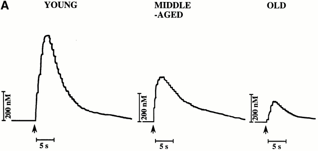
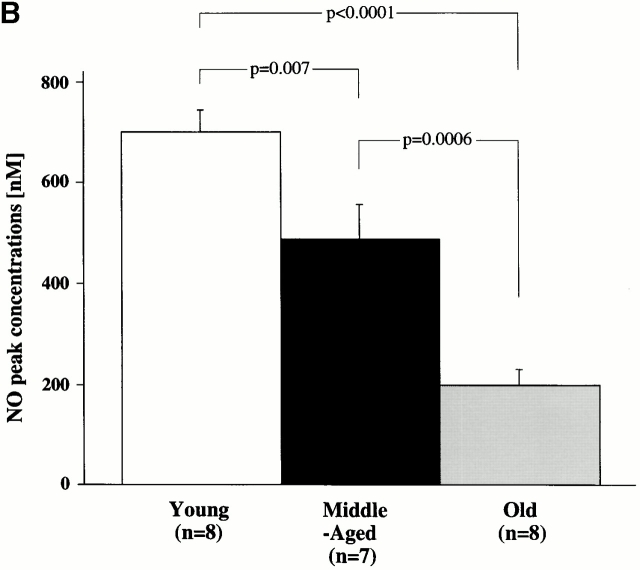
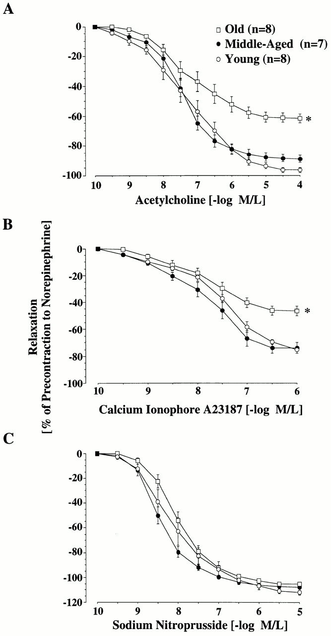
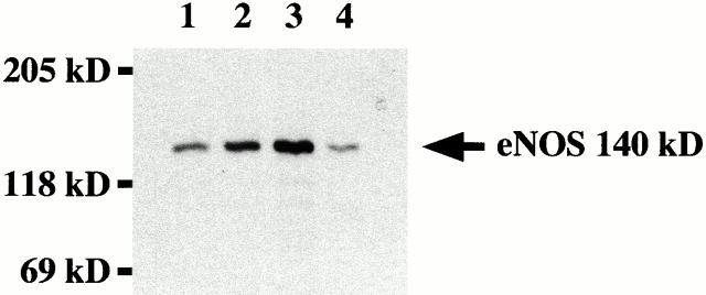
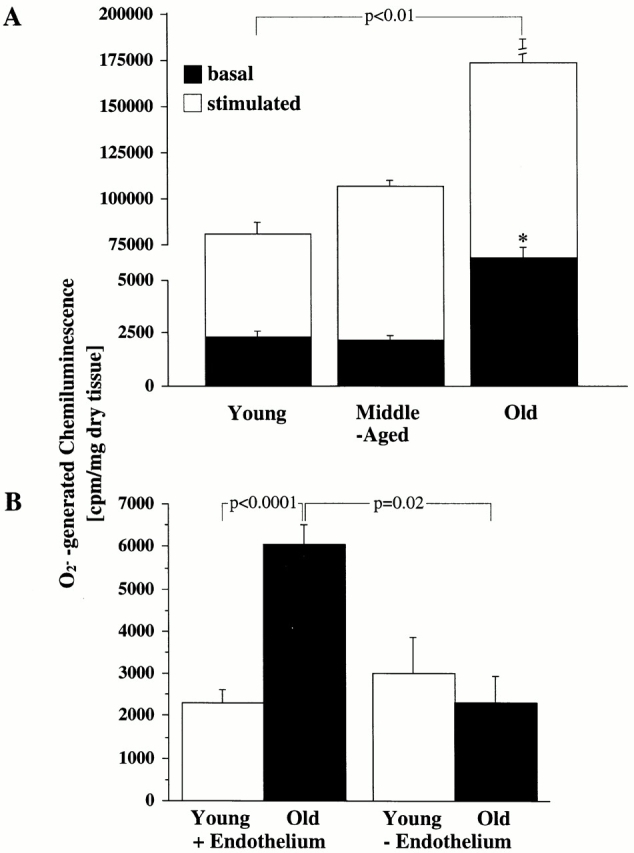
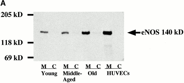
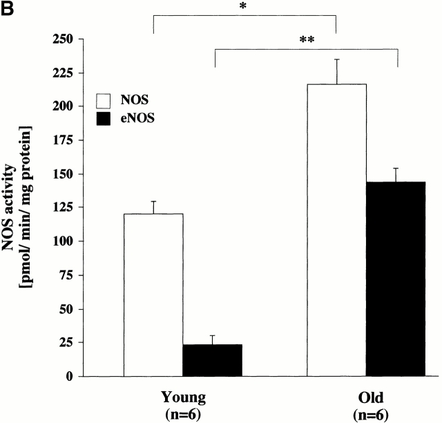
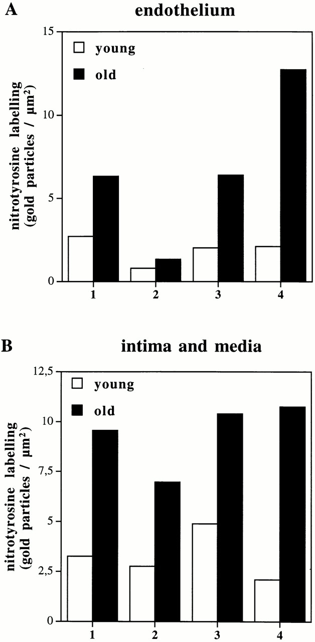
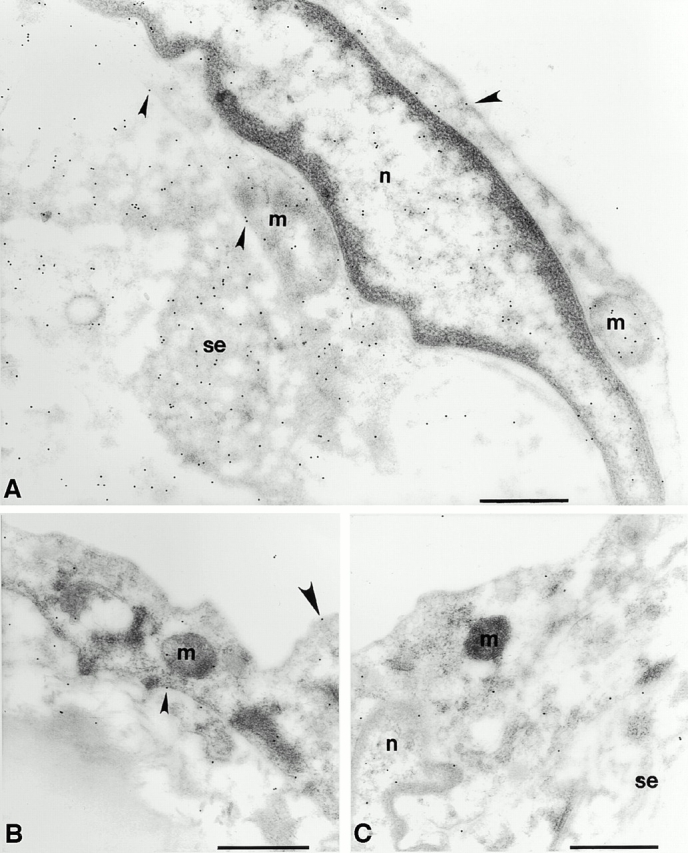
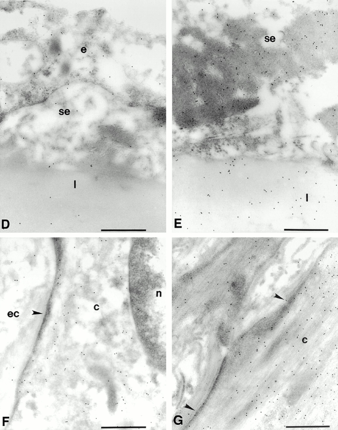
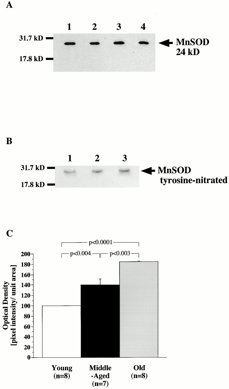
Vascular aging is mainly characterized by endothelial dysfunction. We found decreased free nitric oxide (NO) levels in aged rat aortas, in conjunction with a sevenfold higher expression and activity of endothelial NO synthase (eNOS). This is shown to be a consequence of age-associated enhanced superoxide (.O(2)(-)) production with concomitant quenching of NO by the formation of peroxynitrite leading to nitrotyrosilation of mitochondrial manganese superoxide dismutase (MnSOD), a molecular footprint of increased peroxynitrite levels, which also increased with age. Thus, vascular aging appears to be initiated by augmented.O(2)(-) release, trapping of vasorelaxant NO, and subsequent peroxynitrite formation, followed by the nitration and inhibition of MnSOD. Increased eNOS expression and activity is a compensatory, but eventually futile, mechanism to counter regulate the loss of NO. The ultrastructural distribution of 3-nitrotyrosyl suggests that mitochondrial dysfunction plays a major role in the vascular aging process.


Figures

Figure 2
Figure 2
Age-dependent changes in aortic NO release. (A) Representative amperograms of NO release from isolated aortas. NO release was induced by A23187 (10−6 mol/liter) and measured in situ on the endothelial surface using a porphyrinic microsensor. (B) Bar graph showing peak concentrations of NO.
Figure 2
Figure 2
Age-dependent changes in aortic NO release. (A) Representative amperograms of NO release from isolated aortas. NO release was induced by A23187 (10−6 mol/liter) and measured in situ on the endothelial surface using a porphyrinic microsensor. (B) Bar graph showing peak concentrations of NO.
Figure 1
Figure 1
Age-dependent changes in endothelium-dependent and -independent relaxation of the rat aorta. Line graphs showing concentration–response curves to acetylcholine (A), calcium ionophore A23187 (B), and SNP (C). Relaxation to both endothelium-dependent agonists was reduced in old aortas (P < 0.0001). Endothelium-independent relaxation was unaffected by age. M/L, mol/liter.
Figure 3
Figure 3
eNOS expression in rat aorta. Homogenates of aortic endothelium from young (lane 1), middle-aged (lane 2), and old (lane 3) rats were separated by SDS-PAGE and analyzed by Western blotting for eNOS expression. The position of the molecular mass markers is indicated (expressed in kD). Human umbilical vein endothelial cells (lane 4) were used as positive control.
Figure 5
Figure 5
Age-dependent ·O2 production. (A) Bar graph showing both basal amount of chemiluminescence generated by ·O2 in aorta from young, middle-aged, and old rats (*P < 0.0001 versus basal values for young and middle-aged rats) and maximal generation of chemiluminescence after stimulation with calcium ionophore A23187 (10−6 mol/liter). (B) The influence of endothelium: aortic rings from young and old (n = 6 each) rats were mechanically denuded, and the basal chemiluminescence signal was compared with that from intact aortas.
Figure 4
Figure 4
(A) Subcellular analysis of eNOS as detected by immunoblotting. Membrane-associated (M) and cytosolic (C) eNOS expression. Cytosolic and membrane fractions were prepared from homogenates of each age group and analyzed by immunoblotting. Data are representative of three experiments. HUVECs, human umbilical vein endothelial cells. (B) Activity of total NOS and eNOS in aortas from young and old rats. Activity was measured in homogenates of aortic tissue by determination of the rate of conversion of l-[14C]arginine to l-[14C]citrulline. Significance: total NOS, *P < 0.01; eNOS, **P < 0.001.
Figure 4
Figure 4
(A) Subcellular analysis of eNOS as detected by immunoblotting. Membrane-associated (M) and cytosolic (C) eNOS expression. Cytosolic and membrane fractions were prepared from homogenates of each age group and analyzed by immunoblotting. Data are representative of three experiments. HUVECs, human umbilical vein endothelial cells. (B) Activity of total NOS and eNOS in aortas from young and old rats. Activity was measured in homogenates of aortic tissue by determination of the rate of conversion of l-[14C]arginine to l-[14C]citrulline. Significance: total NOS, *P < 0.01; eNOS, **P < 0.001.
Figure 7
Figure 7
Quantitative analysis of immunogold labeling for 3-nitrotyrosines in young and old aortas in endothelium (A) and intima/media (B). Two sections were prepared from randomly selected areas of each aorta (see Materials and Methods). On each section, 15 fields of endothelium, intima, and media were randomly chosen. In each area of endothelium, immunogold labeling density was estimated over heterochromatin 1, euchromatin 2, cytosol 3, and mitochondria 4. In the intima, immunogold labeling was also estimated over the subendothelial space 1 and the elastic lamellae 2. In the media, immunogold labeling density was estimated over the cytosol 3 and mitochondria 4. All data are presented as means of the labeling density in each region of each of two sections from each aorta.
Figure 6
Figure 6
The accumulation of 3-nitrotyrosine is increased in the aorta of the old rat compared with that of the young rat. Representative electron micrographs show the pattern of immunogold labeling for 3-nitrotyrosine in thin sections of young (B, D, and F) and old (A, E, and G) aortas. Primary antibody binding sites were visualized with goat anti–mouse IgG conjugated to 10-nm gold particles. (A) Intima of the aorta from an old rat. Label is densest over mitochondria (m) and strong over nucleoplasm (n) and over endothelial cell cytoplasm. Sparse labeling is associated with the luminal plasmalemma (large arrowheads) and stronger label is present over the abluminal plasmalemma (small arrowheads). Strong labeling is seen in the subendothelial space (se). (B) Intima of the aorta from a young rat. Label is lower over mitochondria and cytoplasm and sparse over the luminal plasmalemma (large arrowhead) and the abluminal plasmalemma (small arrowhead). (C) Intima of an old rat. The primary antisera against nitrotyrosine was preincubated with 20 μmol/liter nitrotyrosine for 1 h before labeling as in A and B. Label density is reduced to levels lower than those seen in the young rats in all compartments. (D) Intima of a young rat showing low levels of labeling over the endothelium (e) and sparse labeling over the subendothelial space and the first elastic lamellae (l). (E) Subendothelial space and the first elastic lamellae of an old rat. Labeling density is much greater in both compartments and is particularly dense over aggregates of electron dense material seen in the subendothelial space. (F) Smooth muscle cell in the medial layer of the aorta of a young rat. Label is strongest over the cytoplasm (c) and low over the nucleoplasm and extracellular space (ec) and seldom seen over the sarcolemma (arrowhead). (G) Smooth muscle cell in the medial layer of the aorta from an old rat. Labeling is much stronger over the cytoplasm and is frequently seen over the sarcolemma (arrowheads). Bars, 0.5 μm. Original magnifications: ×22,000.
Figure 6
Figure 6
The accumulation of 3-nitrotyrosine is increased in the aorta of the old rat compared with that of the young rat. Representative electron micrographs show the pattern of immunogold labeling for 3-nitrotyrosine in thin sections of young (B, D, and F) and old (A, E, and G) aortas. Primary antibody binding sites were visualized with goat anti–mouse IgG conjugated to 10-nm gold particles. (A) Intima of the aorta from an old rat. Label is densest over mitochondria (m) and strong over nucleoplasm (n) and over endothelial cell cytoplasm. Sparse labeling is associated with the luminal plasmalemma (large arrowheads) and stronger label is present over the abluminal plasmalemma (small arrowheads). Strong labeling is seen in the subendothelial space (se). (B) Intima of the aorta from a young rat. Label is lower over mitochondria and cytoplasm and sparse over the luminal plasmalemma (large arrowhead) and the abluminal plasmalemma (small arrowhead). (C) Intima of an old rat. The primary antisera against nitrotyrosine was preincubated with 20 μmol/liter nitrotyrosine for 1 h before labeling as in A and B. Label density is reduced to levels lower than those seen in the young rats in all compartments. (D) Intima of a young rat showing low levels of labeling over the endothelium (e) and sparse labeling over the subendothelial space and the first elastic lamellae (l). (E) Subendothelial space and the first elastic lamellae of an old rat. Labeling density is much greater in both compartments and is particularly dense over aggregates of electron dense material seen in the subendothelial space. (F) Smooth muscle cell in the medial layer of the aorta of a young rat. Label is strongest over the cytoplasm (c) and low over the nucleoplasm and extracellular space (ec) and seldom seen over the sarcolemma (arrowhead). (G) Smooth muscle cell in the medial layer of the aorta from an old rat. Labeling is much stronger over the cytoplasm and is frequently seen over the sarcolemma (arrowheads). Bars, 0.5 μm. Original magnifications: ×22,000.
Figure 8
Figure 8
Total protein expression and age-associated increased nitration of mitochondrial MnSOD. Western blot of MnSOD (A) from young (lane 1), middle-aged (lane 2), and old (lane 3) aortas. Extracted proteins were separated on 15% SDS-PAGE gels and examined by Western blot with a polyclonal antibody against MnSOD. Recombinant MnSOD served as a positive control (lane 4). Molecular weight markers are indicated (in kD) on the left. (B) Nitrotyrosine-containing immunoprecipitated MnSOD from young (lane 1), middle-old (lane 2), and old (lane 3) rats were separated by 15% SDS-PAGE gels and analyzed by Western blot with a polyclonal antibody against nitrotyrosine. Films were exposed for 15 s to 3 min. (C) Quantitative analysis of tyrosine-nitrated MnSOD.

References

    1. Lüscher T.F., Noll G. The endothelium in coronary vascular control. Heart Dis. 1995;3:1–10.
    1. Palmer R.M., Ashton D.S., Moncada S. Vascular endothelial cells synthesize nitric oxide from L-arginine. Nature. 1988;333:664–666. - PubMed
    1. Moncada S., Higgs A. The L-arginine-nitric oxide pathway. N. Engl. J. Med. 1993;329:2002–2012. - PubMed
    1. Moncada S. The 1991 Ulf von Euler Lecture. The L-argininenitric oxide pathway. Acta Physiol. Scand. 1992;145:201–227. - PubMed
    1. Pollock J.S., Förstermann U., Mitchell J.A., Warner T.D., Schmidt H.H.W., Nakane M., Murad F. Purification and characterization of particulate endothelium-derived relaxing factor synthase from cultured and native bovine aortic endothelial cells. Proc. Natl. Acad. Sci. USA. 1991;88:10480–10484. - PMC - PubMed

Publication types

MeSH terms