Phosphorylated peptides are naturally processed and presented by major histocompatibility complex class I molecules in vivo
- PMID: 11120772
- PMCID: PMC2213507
- DOI: 10.1084/jem.192.12.1755
Phosphorylated peptides are naturally processed and presented by major histocompatibility complex class I molecules in vivo
Abstract
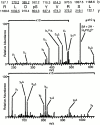
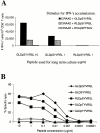
Posttranslational modification of peptide antigens has been shown to alter the ability of T cells to recognize major histocompatibility complex (MHC) class I-restricted peptides. However, the existence and origin of naturally processed phosphorylated peptides presented by MHC class I molecules have not been explored. By using mass spectrometry, significant numbers of naturally processed phosphorylated peptides were detected in association with several human MHC class I molecules. In addition, CD8(+) T cells could be generated that specifically recognized a phosphorylated epitope. Thus, phosphorylated peptides are part of the repertoire of antigens available for recognition by T cells in vivo.
Figures


Similar articles
-
Phosphorylated peptides can be transported by TAP molecules, presented by class I MHC molecules, and recognized by phosphopeptide-specific CTL.J Immunol. 1999 Oct 1;163(7):3812-8. J Immunol. 1999. PMID: 10490979
-
Invariant chain as a vehicle to load antigenic peptides on human MHC class I for cytotoxic T-cell activation.Eur J Immunol. 2014 Mar;44(3):774-84. doi: 10.1002/eji.201343671. Epub 2013 Dec 27. Eur J Immunol. 2014. PMID: 24293164
-
Immunization with a peptide containing MHC class I and II epitopes derived from the tumor antigen SIM2 induces an effective CD4 and CD8 T-cell response.PLoS One. 2014 Apr 1;9(4):e93231. doi: 10.1371/journal.pone.0093231. eCollection 2014. PLoS One. 2014. PMID: 24690990 Free PMC article.
-
Post-Translational Peptide Splicing and T Cell Responses.Trends Immunol. 2017 Dec;38(12):904-915. doi: 10.1016/j.it.2017.07.011. Epub 2017 Aug 19. Trends Immunol. 2017. PMID: 28830734 Review.
-
The other Janus face of Qa-1 and HLA-E: diverse peptide repertoires in times of stress.Microbes Infect. 2010 Nov;12(12-13):910-8. doi: 10.1016/j.micinf.2010.07.011. Epub 2010 Jul 27. Microbes Infect. 2010. PMID: 20670688 Review.
Cited by
-
Recent advances and future perspectives of CAR-T cell therapy in head and neck cancer.Front Immunol. 2023 Jun 29;14:1213716. doi: 10.3389/fimmu.2023.1213716. eCollection 2023. Front Immunol. 2023. PMID: 37457699 Free PMC article. Review.
-
Peptide vaccines in cancer-old concept revisited.Curr Opin Immunol. 2017 Apr;45:1-7. doi: 10.1016/j.coi.2016.11.001. Epub 2016 Dec 9. Curr Opin Immunol. 2017. PMID: 27940327 Free PMC article. Review.
-
Novel Antigen Targets for Immunotherapy of Acute Myeloid Leukemia.Curr Drug Targets. 2017;18(3):296-303. doi: 10.2174/1389450116666150223120005. Curr Drug Targets. 2017. PMID: 25706110 Free PMC article. Review.
-
HLA Class II Presentation Is Specifically Altered at Elevated Temperatures in the B-Lymphoblastic Cell Line JY.Mol Cell Proteomics. 2021;20:100089. doi: 10.1016/j.mcpro.2021.100089. Epub 2021 Apr 29. Mol Cell Proteomics. 2021. PMID: 33933681 Free PMC article.
-
The landscape of MHC-presented phosphopeptides yields actionable shared tumor antigens for cancer immunotherapy across multiple HLA alleles.bioRxiv [Preprint]. 2023 Feb 12:2023.02.08.527552. doi: 10.1101/2023.02.08.527552. bioRxiv. 2023. Update in: J Immunother Cancer. 2023 Sep;11(9):e006889. doi: 10.1136/jitc-2023-006889. PMID: 36798179 Free PMC article. Updated. Preprint.
References
-
- Germain R.N., Margulies D.H. The biochemistry and cell biology of antigen processing and presentation. Annu. Rev. Immunol. 1993;11:403–450. - PubMed
-
- Cresswell P., Bangia N., Dick T., Diedrich G. The nature of the MHC class I peptide loading complex. Immunol. Rev. 1999;172:21–28. - PubMed
-
- Engelhard V.H. Structure of peptides associated with MHC class I molecules. Curr. Opin. Immunol. 1994;6:13–23. - PubMed
-
- Skipper J.C., Gulden P.H., Hendrickson R.C., Harthun N., Caldwell J.A., Shabanowitz J., Engelhard V.H., Hunt D.F., Slingluff C.L., Jr. Mass-spectrometric evaluation of HLA-A*0201-associated peptides identifies dominant naturally processed forms of CTL epitopes from MART-1 and gp100. Int. J. Cancer. 1999;82:669–677. - PubMed
-
- Hunt D.F., Henderson R.A., Shabanowitz J., Sakaguchi K., Michel H., Sevilir N., Cox A.L., Appella E., Engelhard V.H. Characterization of peptides bound to the class I MHC molecule HLA-A2.1 by mass spectrometry. Science. 1992;255:1261–1264. - PubMed
Publication types
MeSH terms
Substances
Grants and funding
LinkOut - more resources
Full Text Sources
Other Literature Sources
Molecular Biology Databases
Research Materials

