

Cracks in the beta-can: fluorescent proteins from Anemonia sulcata (Anthozoa, Actinaria)
- PMID: 11121018
- PMCID: PMC18876
- DOI: 10.1073/pnas.97.26.14091
Cracks in the beta-can: fluorescent proteins from Anemonia sulcata (Anthozoa, Actinaria)
Erratum in
- Proc Natl Acad Sci U S A 2002 Oct 1;99(20):13358
Abstract
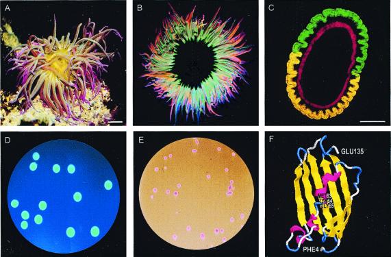
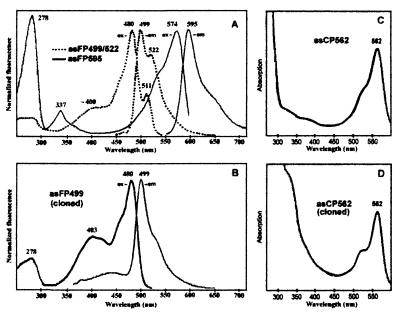
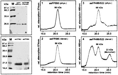
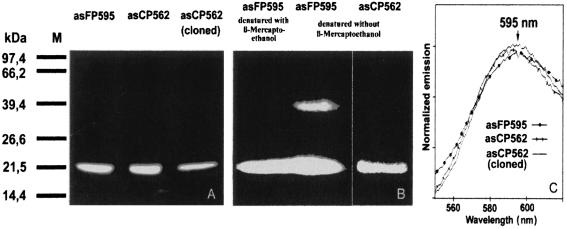
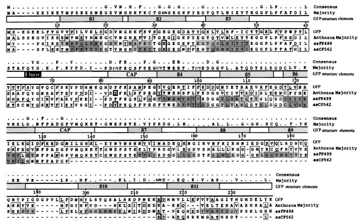
We characterize two green fluorescent proteins (GFPs), an orange fluorescent protein, and a nonfluorescent red protein isolated from the sea anemone Anemonia sulcata. The orange fluorescent protein and the red protein seem to represent two different states of the same protein. Furthermore, we describe the cloning of a GFP and a nonfluorescent red protein. Both proteins are homologous to the GFP from Aequorea victoria. The red protein is significantly smaller than other GFP homologues, and the formation of a closed GFP-like beta-can is not possible. Nevertheless, the primary structure of the red protein carries all features necessary for orange fluorescence. We discuss a type of beta-can that could be formed in a multimerization process.
Figures
Similar articles
-
Natural animal coloration can Be determined by a nonfluorescent green fluorescent protein homolog.J Biol Chem. 2000 Aug 25;275(34):25879-82. doi: 10.1074/jbc.C000338200. J Biol Chem. 2000. PMID: 10852900
-
Variations on the GFP chromophore: A polypeptide fragmentation within the chromophore revealed in the 2.1-A crystal structure of a nonfluorescent chromoprotein from Anemonia sulcata.J Biol Chem. 2005 Jan 28;280(4):2401-4. doi: 10.1074/jbc.C400484200. Epub 2004 Nov 12. J Biol Chem. 2005. PMID: 15542608
-
Traditional GFP-type cyclization and unexpected fragmentation site in a purple chromoprotein from Anemonia sulcata, asFP595.Biochemistry. 2004 Oct 26;43(42):13598-603. doi: 10.1021/bi0488247. Biochemistry. 2004. PMID: 15491166
-
Fluorescent proteins as biomarkers and biosensors: throwing color lights on molecular and cellular processes.Curr Protein Pept Sci. 2008 Aug;9(4):338-69. doi: 10.2174/138920308785132668. Curr Protein Pept Sci. 2008. PMID: 18691124 Free PMC article. Review.
-
Green fluorescent protein.Photochem Photobiol. 1995 Oct;62(4):651-6. doi: 10.1111/j.1751-1097.1995.tb08712.x. Photochem Photobiol. 1995. PMID: 7480149 Review.
Cited by
-
The mitochondrial 60-kDa heat shock protein in marine invertebrates: biochemical purification and molecular characterization.Cell Stress Chaperones. 2004 Mar;9(1):38-48. doi: 10.1379/469.1. Cell Stress Chaperones. 2004. PMID: 15270076 Free PMC article.
-
Toxicity Profiling of Bacterial Inclusion Bodies in Human Caco-2 Cells.Front Bioeng Biotechnol. 2022 Apr 29;10:842256. doi: 10.3389/fbioe.2022.842256. eCollection 2022. Front Bioeng Biotechnol. 2022. PMID: 35573225 Free PMC article.
-
Fluorescent proteins of the EosFP clade: intriguing marker tools with multiple photoactivation modes for advanced microscopy.RSC Chem Biol. 2021 Mar 31;2(3):796-814. doi: 10.1039/d1cb00014d. eCollection 2021 Jun 1. RSC Chem Biol. 2021. PMID: 34458811 Free PMC article. Review.
-
Identification of GFP-like proteins in nonbioluminescent, azooxanthellate anthozoa opens new perspectives for bioprospecting.Mar Biotechnol (NY). 2004 May-Jun;6(3):270-7. doi: 10.1007/s10126-004-3006-4. Epub 2004 May 13. Mar Biotechnol (NY). 2004. PMID: 15136917
-
Coexistence of nonfluorescent chromoproteins and fluorescent proteins in massive Porites spp. corals manifesting a pink pigmentation response.Front Physiol. 2024 Jun 17;15:1339907. doi: 10.3389/fphys.2024.1339907. eCollection 2024. Front Physiol. 2024. PMID: 38952870 Free PMC article.
References
-
- Shimomura O, Johnson F H, Saiga Y. Aequorea J Cell Comp Physiol. 1962;59:223–239. - PubMed
-
- Morin J G, Hastings J W. J Cell Physiol. 1971;77:305–311. - PubMed
-
- Wampler J E, Hori K, Lee J W, Cormier M J. Renilla Biochem. 1971;10:2903–2909. - PubMed
-
- Wampler J E, Karkhanis Y D, Morin J G, Cormier M J. Biochim Biophys Acta. 1973;314:104–109. - PubMed
-
- Cormier M J, Hori K, Anderson J M. Biochim Biophys Acta. 1974;364:137–164. - PubMed
MeSH terms
Substances
Associated data
- Actions
- Actions
LinkOut - more resources
Full Text Sources
Other Literature Sources
