. 2000 Dec 19;97(26):14301-6.
doi: 10.1073/pnas.97.26.14301.

c-Mos forces the mitotic cell cycle to undergo meiosis II to produce haploid gametes

Affiliations

c-Mos forces the mitotic cell cycle to undergo meiosis II to produce haploid gametes

K Tachibana et al. Proc Natl Acad Sci U S A. .

Abstract

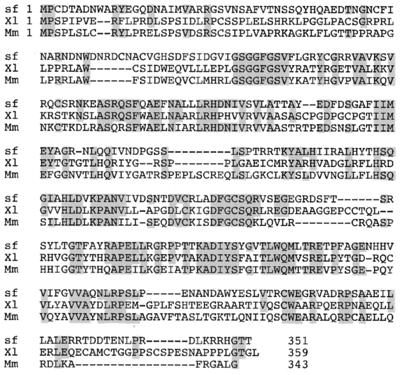
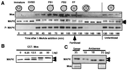
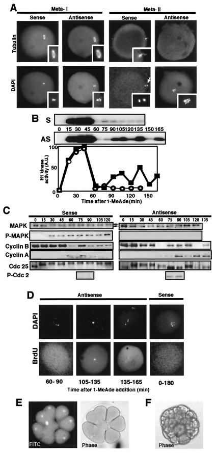
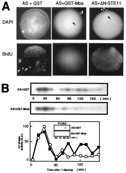
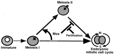
The meiotic cycle reduces ploidy through two consecutive M phases, meiosis I and meiosis II, without an intervening S phase. To maintain ploidy through successive generations, meiosis must be followed by mitosis after the recovery of diploidy by fertilization. However, the coordination from meiotic to mitotic cycle is still unclear. Mos, the c-mos protooncogene product, is a key regulator of meiosis in vertebrates. In contrast to the previous observation that Mos functions only in vertebrate oocytes that arrest at meiotic metaphase II, here we isolate the first invertebrate mos from starfish and show that Mos functions also in starfish oocytes that arrest after the completion of meiosis II but not at metaphase II. In the absence of Mos, meiosis I is followed directly by repeated embryonic mitotic cycles, and its reinstatement restores meiosis II and subsequent cell cycle arrest. These observations imply that after meiosis I, oocytes have a competence to progress through the embryonic mitotic cycle, but that Mos diverts the cell cycle to execute meiosis II and remains to restrain the return to the mitotic cycle. We propose that a role of Mos that is conserved in invertebrate and vertebrate oocytes is not to support metaphase II arrest but to prevent the meiotic/mitotic conversion after meiosis I until fertilization, directing meiosis II to ensure the reduction of ploidy.


Figures

Figure 1
Figure 1
Deduced amino acid sequence of starfish Mos and its alignment with mouse and Xenopus Mos proteins. Identical amino acids are shaded, and gaps introduced for optimal alignment are indicated by dashes. The starfish (sf) Mos sequence has been deposited in DNA Data Base of Japan/European Molecular Biology Laboratory/GenBank under accession no. AB040102. Mm, mouse (accession no. J00372); Xl, Xenopus laevis (accession no. X13311).
Figure 2
Figure 2
Mos functions as an MAP kinase activator in starfish oocytes. (A) Dynamics of Mos and MAP kinase through starfish meiotic cycles. At the female pronucleus (FP) stage after the completion of meiosis II, mature eggs were fertilized, resulting in the disappearance of Mos. P-MAPK, antiphospho MAP kinase corresponding to its active form; PB 1 and 2, the first and the second polar bodies, respectively. (B) MAP kinase activation induced by injection of the GST-starfish Mos fusion protein into immature starfish oocytes. Oocytes were injected with various amounts of GST-Mos and recovered at 30 min for immunoblots. (C) Antisense mos prevents MAP kinase activation after 1-MeAde addition in starfish oocytes. Immature oocytes were injected with various amounts of antisense and sense mos oligonucleotides and then treated with 1-MeAde to undergo GVBD. Oocytes were recovered for immunoblots 60 min after 1-MeAde addition. Mos synthesis was undetectable at 35 pg injection of antisense mos. Upper and lower bands of MAP kinase (indicated by arrows) correspond to the active and the inactive forms, respectively (see ref. 19).
Figure 3
Figure 3
The mitotic type of cell cycles proceed after meiosis I in Mos-deficient 1-MeAde-treated unfertilized starfish oocytes. (A) Abortive spindle formation at a stage corresponding to metaII in Mos-deficient oocytes. After 1-MeAde addition to oocytes that had been injected with either antisense or sense mos, a spindle was frequently detectable at metaI with antitubulin staining but thereafter could not be detected, and condensed chromosomes were located in the middle of each oocyte. (Insets) Meiotic spindles and condensed chromosomes at higher magnification. White arrow indicates the first polar body. (B) Repeated fluctuation of histone H1 kinase activity after 1-MeAde addition in starfish oocytes injected with antisense mos. (Upper) Autoradiograms (AS, antisense mos; S, sense mos). (Lower) Radioactivity of the excised histone H1 bands (closed squares, antisense mos; open circles, sense mos). (C) Dynamics of cyclins B and A, Cdc25, Tyr phosphorylation in Cdc2 and MAP kinase after 1-MeAde addition in starfish oocytes injected with antisense mos. P-MAPK and P-Cdc2, immunoblots with antiphospho MAP kinase and antiphospho-Tyr-15 of Cdc2, respectively. (D) DNA replication occurs when histone H1 kinase activity drops to minimal levels after 1-MeAde addition to starfish oocytes injected with antisense mos. BrdUrd incorporation by pulse labeling for 30 min indicated below each panel was detectable rarely immediately after meiosis I (see also Fig. 4A) and always thereafter in antisense mos-injected oocytes; in contrast, it was undetectable through 180 min continuous labeling in control sense mos-injected oocytes. Each oocyte was double stained with 4′,6-diamidino-2-phenylindole (DAPI). (E) Several rounds of cleavage in antisense mos-injected, 1-MeAde-treated, unfertilized starfish oocytes. Nuclear divisions and furrowing occurred normally in some blastomeres and abnormally in others within an embryo. Thus, although they were abnormal as a whole, these oocytes developed to the two-cell stage almost invariably and to the blastula stage at 10%. Presence of nucleus is shown by accumulation of FITC-conjugated BSA coupled with nucleoplasmin NLS peptide, which was injected into immature oocytes (Left). (F) Development to 64-cell stage embryo in 1-MeAde- and U0126-treated unfertilized starfish oocytes. Almost all of these embryos developed to bipinnaria larvae, even though abnormal cleavages were partially observed. Note the absence of elevation of the fertilization envelope.
Figure 4
Figure 4
Restoration of meiosis II by Mos in starfish oocytes in which translation of mos is prevented. (A) Formation of two polar bodies along with a female pronucleus after 1-MeAde addition to oocytes that had received the injection of antisense mos (AS) and either GST-Mos or ΔN-STE11. Polar bodies (arrowheads) were detected by DAPI staining. Black arrow indicates the female pronucleus. BrdUrd incorporation was undetectable in female pronuclei of GST-Mos or ΔN-STE11-restored oocytes but could be detected in control oocytes (white arrows). (B) Two limited rounds of H1 kinase activity after 1-MeAde addition to oocytes that had received the injection of antisense mos and the GST-Mos fusion protein. After 1-MeAde addition, oocytes were recovered at indicated times and processed for measurement of H1 kinase activity (Top and Middle for autoradiogram; Bottom for radioactivity of the excised histone H1 bands; open circles, injection with antisense mos plus GST-Mos; closed squares, injection with antisense mos plus GST) and for Tyr phosphorylation in Cdc2 (Bottom, Insets). In both A and B, control oocytes had been injected with antisense mos (AS) and GST before 1-MeAde addition.
Figure 5
Figure 5
A role of Mos is conserved in vertebrate and invertebrate oocytes. At the end of meiosis I, the competence to undergo the embryonic mitotic cell cycle is already acquired, but Mos forces the embryonic mitotic cycle to undergo meiosis II, thus enabling the reduction of ploidy. Thereafter, Mos remains to restrain the return to the embryonic mitotic cycle, thus preventing parthenogenetic development. Fertilization resets the Mos-dependent detour of the cell cycle, leading to the recovery of the embryonic mitotic cycle. Thus, Mos is a key coordinator of meiotic/mitotic conversion.

Similar articles

Cited by

References

    1. Sagata N. Trends Cell Biol. 1996;6:22–28. - PubMed
    1. Oskarsson M, McClements W L, Blair D G, Maizel L V, Vande Woude G F. Science. 1980;207:1222–1224. - PubMed
    1. Posada J, Yew N, Ahn N G, Vande Woude G F, Cooper J A. Mol Cell Biol. 1993;13:2546–2553. - PMC - PubMed
    1. Shibuya E K, Ruderman J V. Mol Biol Cell. 1993;4:781–790. - PMC - PubMed
    1. Nebreda A R, Hunt T. EMBO J. 1993;12:1979–1986. - PMC - PubMed

Publication types

Substances

Associated data

LinkOut - more resources