. 2000 Dec 19;97(26):14352-7.
doi: 10.1073/pnas.97.26.14352.

The TACC domain identifies a family of centrosomal proteins that can interact with microtubules

Affiliations

The TACC domain identifies a family of centrosomal proteins that can interact with microtubules

F Gergely et al. Proc Natl Acad Sci U S A. .

Abstract

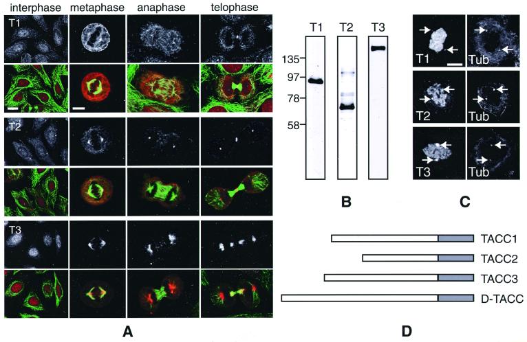
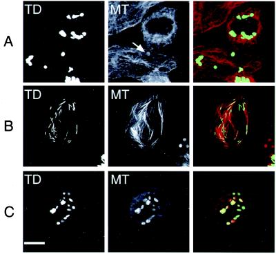
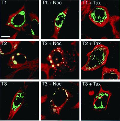
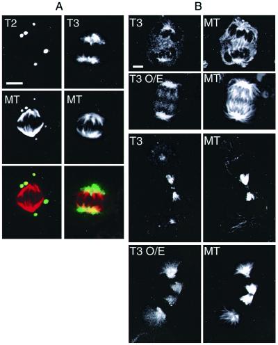
We recently showed that the Drosophila transforming acidic coiled-coil (D-TACC) protein is located in the centrosome, interacts with microtubules, and is required for mitosis in the Drosophila embryo. There are three known human TACC proteins that share a conserved, C-terminal, coiled-coil region with D-TACC. These proteins have all been implicated in cancer, but their normal functions are unknown. We show that all three human TACC proteins are concentrated at centrosomes, but with very different characteristics: TACC1 is weakly concentrated at centrosomes during mitosis; TACC2 is strongly concentrated at centrosomes throughout the cell cycle; and TACC3 is strongly concentrated in a more diffuse region around centrosomes during mitosis. When the C-terminal TACC domain is overexpressed in HeLa cells, it forms large polymers in the cytoplasm that can interact with both microtubules and tubulin. The full-length TACC proteins form similar polymers when overexpressed, but their interaction with microtubules and tubulin is regulated during the cell cycle. At least one of the human TACC proteins appears to increase the number and/or stability of centrosomal microtubules when overexpressed during mitosis. Thus, the TACC domain identifies a family of centrosomal proteins that can interact with microtubules. This may explain the link between the TACC genes and cancer.


Figures

Figure 1
Figure 1
A comparison of the known human TACC proteins. (A) Fixed HeLa cells at interphase, metaphase, anaphase, and telophase were stained with anti-TACC antibodies (Top; red in color panels) and anti-tubulin antibodies (green in color panels). In this panel, and in all subsequent panels, T1 denotes TACC1, T2 denotes TACC2, and T3 denotes TACC3. Scale bars: interphase, 10 μm; mitotic cells, 5 μm. (B) Western blots of HeLa cell extracts probed with affinity-purified anti-TACC1 (lane 1), anti-TACC2 (lane 2), and anti-TACC3 (lane 3) antibodies. (C) Nocodazole-treated cells were stained with the anti-TACC antibodies (Left) and with a mixture of anti-α-tubulin and anti-γ-tubulin antibodies (Right) to monitor the location of the centrosomes and to confirm that microtubules were depolymerized. All of the TACC antibodies stained the centrosomes (arrows) even though there were no visible microtubules in the cell. Note that the chromosomes in these cells were counterstained with propidium iodide, and the signal from this fluorophore “bleeds through” into the channel used to detect the TACC antibodies. (Scale bar = 4 μm.) (D) A schematic diagram of the TACC proteins. The shaded boxes represent the conserved 200-aa TACC domain that is predicted to form a coiled-coil.
Figure 2
Figure 2
The behavior of the overexpressed TACC domains is shown in normal cells (Top) and in cells where microtubules are stabilized by taxol (Middle) or depolymerized by nocodazole (Bottom). The TACC domain is visualized by the fluorescence of the GFP tag (Left). Microtubules were stained with anti-tubulin antibodies (Center). A merged image is shown (Right): in this and all subsequent merged panels, the GFP-TACC fusion protein is shown in green and tubulin in red. The arrow highlights the weak association of tubulin with a TACC domain structure in an untreated cell. In nocodazole-treated cells, the unpolymerized tubulin is concentrated around the TACC domain structures, whereas in taxol the structures stretch out along the microtubule bundles. Only the TACC2 TACC domain is shown, as the TACC domains of TACC1 and TACC3 behave identically. (Scale bar = 10 μm.)
Figure 3
Figure 3
The large TACC-containing structures are highly ordered polymers. (A) Immuno-electron microscopy of cells overexpressing the GFP-TACC domain fusion proteins. Cells were fixed with formaldehyde and stained with anti-GFP antibodies, followed by Nanogold-labeled secondary antibodies and silver intensification. In thin sections, the silver particles stain the edges of large, globular, cytoplasmic structures that were composed of a regularly spaced, electron dense, polymer (the cell shown here is overexpressing the TACC2 TACC domain). The staining is largely confined to the margins of these structures probably because the formaldehyde fixation has highly cross-linked the structures, impeding internal antibody access. A 2× higher magnification view of part of this structure is shown in B. The ordered morphology of these structures is better preserved in glutaraldehyde-fixed cells shown in C and, at 2× higher magnification, in D. This cell is expressing a GFP full-length TACC2 fusion protein, and the largest diameter of this TACC structure is about half the size of this cell's nucleus. (Scale bar = 0.5 μm.)
Figure 4
Figure 4
The behavior of the overexpressed full-length TACC proteins. In normal transfected cells (Left), all of the overexpressed TACC proteins (green) form large structures in the cytoplasm. Tubulin is shown in red in the merged images; when tubulin is concentrated around the TACC structures, they appear to be yellow. Tubulin is not highly concentrated around the TACC1 or TACC3 polymers but is concentrated around the TACC2 polymers. In nocodazole-treated cells (Center), the TACC1 polymers do not interact with tubulin and TACC2 polymers strongly interact with tubulin, whereas the TACC3 polymers weakly interact with tubulin. In taxol-treated cells (Right), only the TACC2 polymers interact with the stabilized microtubules. (Scale bar = 10 μm.)
Figure 5
Figure 5
The behavior of overexpressed TACC2 and TACC3 in mitotic cells. (A) The TACC2 polymers (green in merged image) remain highly compacted throughout mitosis but strongly interact with the unpolymerized tubulin (red in merged image) in the cell. During mitosis, the TACC3 polymers are much less compacted than in interphase, and they are strongly concentrated around the poles of the mitotic spindle. (Scale bar = 5 μm.) (B) TACC3 appears to increase the number of centrosomal microtubules. A comparison between transfected cells overexpressing TACC3 (T3 O/E) and nontransfected cells stained with anti-TACC3 (Left) and anti-tubulin antibodies (Right). In both anaphase (Upper) and telophase (Lower), the centrosomes in TACC3 overexpressing cells appear to be associated with many more microtubules. Note that all of these images were taken from transfected and nontransfected cells on the same coverslip with identical settings on the confocal microscope so that meaningful comparisons could be made between them.

References

    1. Glover D M, Gonzalez C, Raff J W. Sci Am. 1993;268:62–68. - PubMed
    1. Kellogg D R, Moritz M, Alberts B M. Annu Rev Biochem. 1994;63:639–674. - PubMed
    1. Desai A, Mitchison T J. Annu Rev Cell Dev Biol. 1997;13:83–117. - PubMed
    1. Moritz M, Braunfeld M B, Sedat J W, Alberts B, Agard D A. Nature (London) 1995;378:638–640. - PubMed
    1. Zheng Y X, Wong M L, Alberts B, Mitchison T. Nature (London) 1995;378:578–583. - PubMed

Publication types