. 2000 Dec 19;97(26):14506-11.
doi: 10.1073/pnas.97.26.14506.

UL82 virion protein activates expression of immediate early viral genes in human cytomegalovirus-infected cells

Affiliations

UL82 virion protein activates expression of immediate early viral genes in human cytomegalovirus-infected cells

W A Bresnahan et al. Proc Natl Acad Sci U S A. .

Abstract

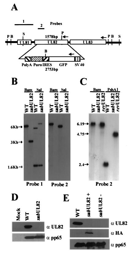
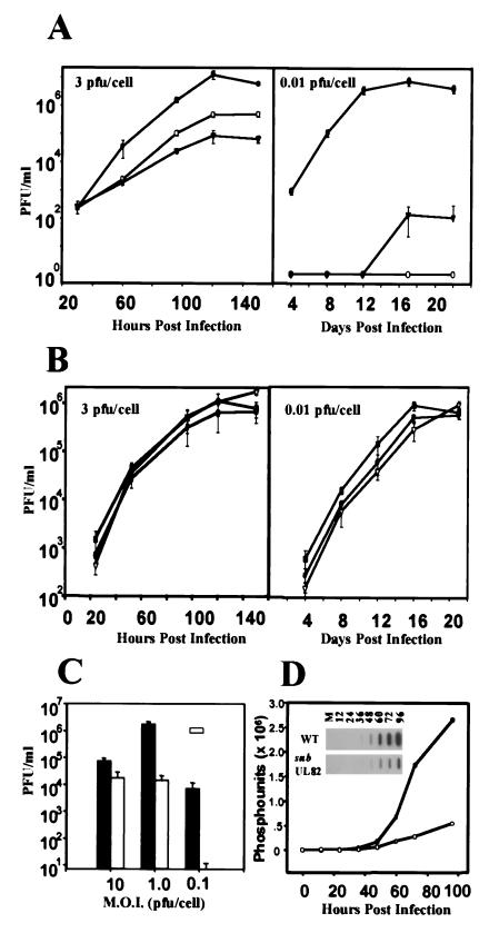
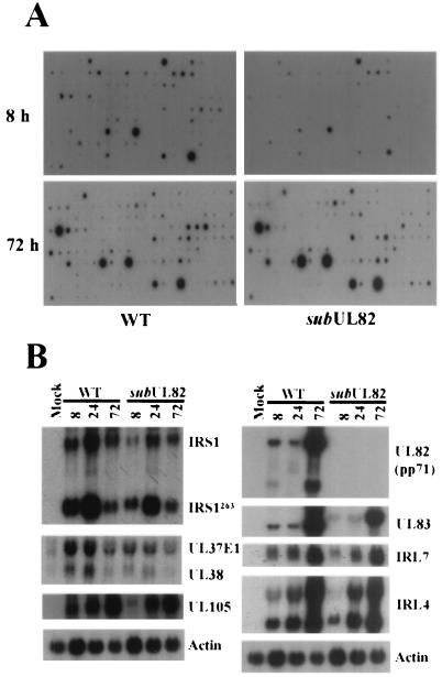
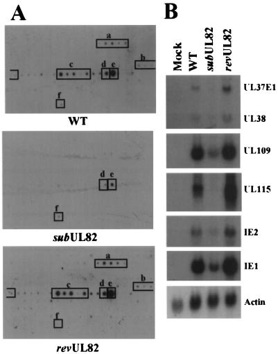
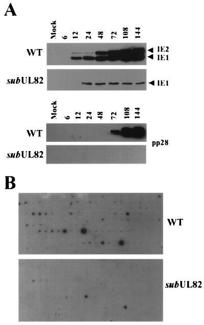
The human cytomegalovirus UL82 gene encodes a protein (pp71) that is localized in the tegument domain of the virus particle. The UL82 gene product is delivered to the nucleus at the time of infection, and it is believed to function in gene activation. We have constructed a human cytomegalovirus mutant, ADsubUL82, that lacks a substantial portion of the UL82 coding region. It was propagated on human diploid fibroblasts expressing the UL82 gene product, and it was possible to produce a mutant virus lacking the UL82 protein by passaging virus stocks for one cycle of growth on normal, noncomplementing fibroblasts. The UL82-deficient mutant displays a multiplicity-dependent growth defect in normal human fibroblasts. The growth of ADsubUL82 is severely restricted at low input multiplicities (0.01-0.1 plaque-forming units per cell), producing a yield that is reduced by a factor of about 10(5) in comparison to wild-type virus. At higher input multiplicities (10 plaque-forming units per cell), ADsubUL82 grew nearly as well as the wild-type virus. By using a human cytomegalovirus gene array, we demonstrated that UL82 functions to facilitate virus mRNA accumulation very early during the human cytomegalovirus replication cycle. The growth phenotype associated with the UL82 mutant seems to result from its inability to efficiently activate human cytomegalovirus immediate early genes.


Figures

Figure 1
Figure 1
Characteristics of ADsubUL82. (A) Schematic representation of the UL81–UL83 region of the HCMV genome, the green fluorescent protein (GFP)/internal ribosomal entry site (IRES)/puromycin (Puro) cassette controlled by the simian virus 40 promoter and poly(A) site substituted for the UL82 ORF, and the probes used to characterize the mutant virus. BamHI (B), SalI (S), and PshAI (P) restriction sites are shown. (B) Southern blot analysis of wt AD169 (WT) and ADsubUL82 (subUL82) viral DNA. Viral DNA was digested with either BamHI or SalI, separated by electrophoresis, transferred to nitrocellulose, and analyzed with 32P-labeled probe 1 or probe 2. (C) Southern blot analysis of wt, ADsubUL82, and ADrevUL82 viral DNA. Viral DNA was digested with either BamHI or PshAI and analyzed with 32P-labeled probe 2, which encompasses a portion of the UL82 ORF. (D) Western blot analysis of UL82 (α UL82) expression at 120 h after infection of human fibroblasts with wt or ADsubUL82. As a control, expression of pp65 (α pp65) was also measured. (E) Detection of UL82 protein packaged within virus particles. Equal amounts of partially purified wt, ADsubUL82+UL82, or ADsubUL82−UL82 virus particles were assayed for the presence of UL82 protein with either a UL82-specific antibody (α UL82) or a hemagglutinin-specific (α HA) antibody directed against an epitope contained within the UL82 protein expressed from WF28-71-HA cells. Incorporation of the tegument protein pp65 (α pp65) was also measured.
Figure 2
Figure 2
Growth kinetics of wt and mutant viruses. (A) Human fibroblasts were infected (3 or 0.01 pfu per cell) with wt (●), ADsubUL82+UL82 (○), or ADsubUL82−UL82 (▾) virus. Cultures were harvested at the indicated times after infection, and infectious virus was quantified by plaque assay on WF28-71-HA cells. (B) Human fibroblasts were infected (3 or 0.01 pfu per cell) with wt (●), ADrevUL82#1 (■), or ADrevUL82#2 (▿) virus. Viruses designated 1 and 2 are two independent isolates of the revertant virus. Cultures were harvested at the indicated times after infection, and virus was quantified by plaque assay on human fibroblasts. (C) Human fibroblasts were infected (10, 1.0, or 0.1 pfu per cell) with wt (black bars) or ADsubUL82−UL82 (open bars) virus. Cultures were harvested at 144 h after infection, and virus was quantified by plaque assay on WF28-71-HA cells. M.O.I., multiplicity of infection. (D) Accumulation of wt and ADsubUL82 viral DNA in human fibroblasts infected at a multiplicity of 2 pfu per cell. DNA was isolated at the indicated times after infection, and viral DNA was quantified by slot blot analysis with an HCMV-specific probe.
Figure 3
Figure 3
HCMV mRNA accumulation after infection with wt or mutant viruses at a relatively high multiplicity. (A) Human fibroblasts were infected with wt or ADsubUL82−UL82 virus (2 pfu per cell). RNA was isolated 8 and 72 h after infection and reverse transcribed in the presence of [32P]dCTP. Quantities of 32P-labeled cDNA copied from RNA that was prepared from the same number of cells infected with wt or mutant virus was used as probe. The arrays contained an actin control DNA that was visible after longer exposure times than are displayed here. The intensity of the actin spot was similar for each preparation of probe. (B) Human fibroblasts were infected with wt or ADsubUL82−UL82 virus (2 pfu per cell). Total RNA was isolated at 8, 24, and 72 h after infection and assayed for HCMV gene expression by Northern blotting with 32P-labeled gene-specific probes. Actin was included as an internal loading control.
Figure 4
Figure 4
HCMV IE mRNA accumulation after infection with wt or mutant viruses in the presence of cycloheximide. (A) Human fibroblasts were infected with wt, ADsubUL82−UL82, or ADrevUL82 virus (1 pfu per cell) in the presence of cycloheximide (100 μg/ml). At 8 h after infection, mRNA was isolated and reverse transcribed in the presence of [32P]dCTP. The labeled cDNA was then used to probe HCMV gene arrays on membranes. Transcripts expressed after infection with the wt or revertant virus correspond to the following open reading frames: UL36–UL38, a; UL106–UL109, b; UL115–UL119, c; UL122, d; UL123, e; and actin, f. Actin was used to confirm that equal quantities of each sample were analyzed, as described in the legend for Fig. 3A. (B) Human fibroblasts were infected with wt, ADsubUL82−UL82, or ADrevUL82 virus (1 pfu per cell) in the presence of cycloheximide (100 μg/ml). Total RNA was isolated at 8 h after infection and assayed for HCMV IE gene expression by Northern blotting with 32P-labeled gene-specific probes.
Figure 5
Figure 5
HCMV mRNA accumulation after infection with wt or mutant viruses at a relatively low multiplicity. (A) Human fibroblasts were infected with wt or ADsubUL82−UL82 virus (0.1 pfu per cell). Cell lysates were prepared at the indicated times after infection and assayed for IE1, IE2, and pp28 protein expression by Western blotting. (B) Human fibroblasts were infected with wt or ADsubUL82−UL82 virus at a multiplicity of 0.1 pfu per cell. At 144 h after infection, RNA was isolated and reverse transcribed in the presence of [32P]dCTP. The labeled cDNA was then used to probe HCMV gene arrays. Actin was used to confirm that equal quantities of each sample were analyzed, as described in the legend for Fig. 3A.

References

    1. Alford C A, Britt W J. In: Fields Virology. Fields B N, Knipe D M, Howely P M, editors. New York: Lippincott-Raven; 1995. pp. 2493–2543.
    1. Baldick C J, Jr, Shenk T. J Virol. 1996;70:6097–6105. - PMC - PubMed
    1. Gibson W. Virology. 1981;111:516–537. - PubMed
    1. Gibson W. Virology. 1983;128:391–406. - PubMed
    1. Spaete R R, Gehrz R C, Landini M P. J Gen Virol. 1994;75:3287–3308. - PubMed

Publication types

LinkOut - more resources