. 2000 Dec 19;97(26):14566-71.
doi: 10.1073/pnas.97.26.14566.

Highly restricted spread of HIV-1 and multiply infected cells within splenic germinal centers

Affiliations

Highly restricted spread of HIV-1 and multiply infected cells within splenic germinal centers

S Gratton et al. Proc Natl Acad Sci U S A. .

Abstract

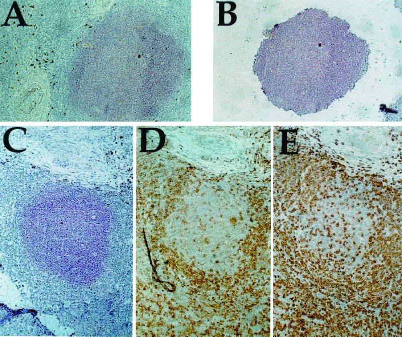
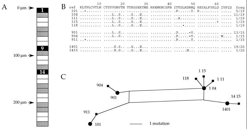
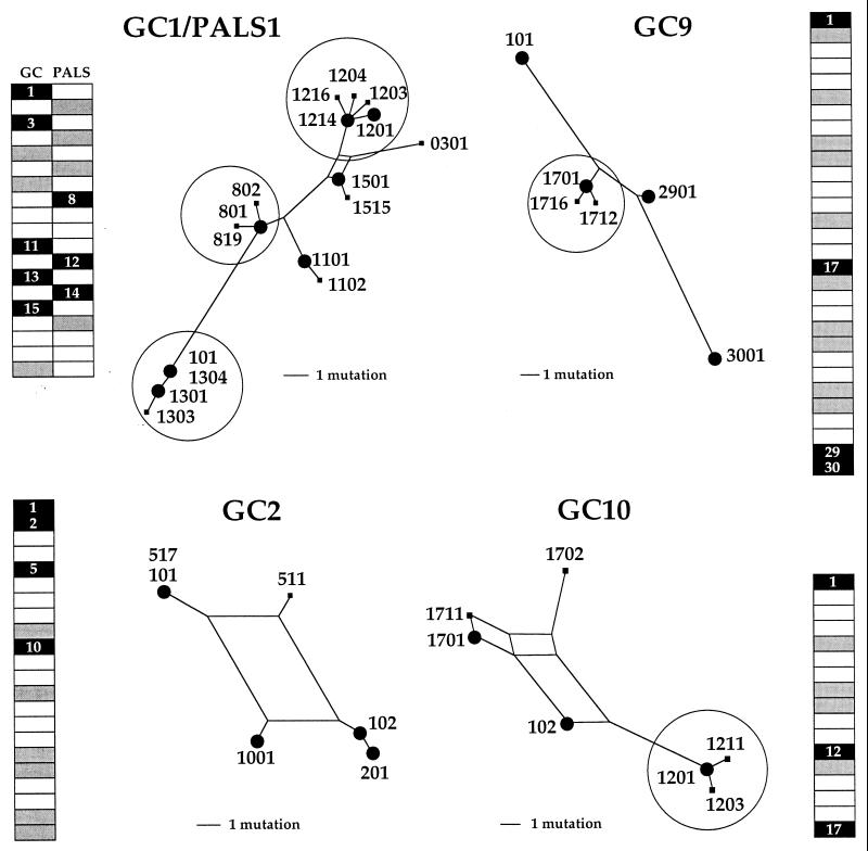
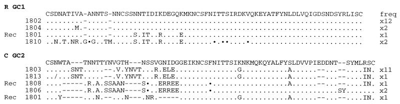
The tremendous dynamics of HIV infection finds expression in the tempo of sequence diversification. Genetic diversity calculations require the clearance of a majority of infected cells, the obvious predator being anti-HIV immune responses. Indeed, infiltration of germinal centers (GCs) by HIV-specific CD8(+) cytotoxic T lymphocytes has been described. A corollary to this description would be limited diffusion of virus within lymphoid structures. HIV efficiently infects and replicates mainly in activated CD4(+) T lymphoblasts. These cells are found within GCs after their activation in the adjacent periarteriolar lymphoid sheath (PALS). Here GCs and PALS have been dissected from consecutive 10-micrometer sections through splenic tissue from three HIV-1-infected patients. Nested PCR amplification of the two first hypervariable regions of the env gene indicated that 38-78% of sections contained HIV-infected cells. Since there are several hundred CD4(+) T cells per GC section, approximately 0.09-0.64% harbor proviral DNA. Such a low frequency not only suggests that virions on the follicular dendritic cell surfaces do not readily infect adjacent T cells but also indicates highly restricted spread of HIV within GCs and the PALS. Sections were heavily infiltrated by CD8(+) cells, which, together with a large body of extant data, suggests that the majority of infected cells are destroyed by HIV-specific cytotoxic T lymphocytes before becoming productively infected. Finally, sequence analysis revealed that those HIV-positive cells were multiply infected, which helps explain widespread recombination despite a low overall frequency of infected cells.


Figures

Figure 1
Figure 1
Tissue sections were treated as described in the text. Here are shown sections from patient R. (A) DRC-1-stained tissue before dissection. (B) The same GC after dissection, showing the amount of tissue picked up for analysis. (C–E) DRC-1 (C), CD4 (D), and CD8 (E) stained GC. (×10.)
Figure 2
Figure 2
Spatial distribution of HIV DNA, sequences, and phylogenetic analysis from patient A-GC-6. (A) The GC is represented as a long rectangle, boxes representing the different sections. Black and gray indicate HIV-1 DNA-positive and -negative sections, respectively. The white sections in between were stained for CD4, CD3, or CD8. Sections are numbered from top to bottom. The vertical scale is given in micrometers. (B) Sequences present in the HIV-positive cuts. All sequences were aligned to that of an internal reference. Only sequence differences are shown; ⋅s represent synonymous substitutions; and frequencies (freq) of the sequences are given on the right. The sequence code is appended to the section number. For example, 901 refers to sequence 01 from section 9. (C) Phylogenic relationship of A-GC6 sequences determined by the SplitsTree2 program. The distances separating sequences in the trees are strictly proportional to the number of mutations: a single substitution, insertion, or deletion.
Figure 3
Figure 3
Spatial distribution of HIV DNA and phylogenetic analysis of sequences from four GCs from patient A. Three GCS and a GC/PALS pair from patient A were analyzed. They are represented by long rectangles at the periphery. Black and gray indicate HIV DNA-positive and -negative sections, respectively. White sections in between were stained for CD4, CD3, or CD8. Phylogenetic analyses corresponding to HIV-positive sections are presented toward the center of the figure. Clusters of sequences differing by a single event are encircled. Large dots indicate a sequence found at a frequency of >20% of all those derived from the section. For the record, one sequence (GC9–3001) is a defective G → A hypermutant, demonstrating that it was amplified from a single cell, as there was no other sequence in the section that could play the role of helper virus. It also reiterates the sensitivity of env V1V2 nested PCR. A few identical sequences were found in different GCs, notably: GC2-101 and GC6-101; GC2-102 and GC6-104; GC2-511 and GC6-901; and GC3-602 and GC6-911.
Figure 4
Figure 4
Recombinants within GC sections. Recombinant genome (Rec 1801) within GC1 section 18 from patient R and a collection of recombinants (Recs 1808 and 1801) found within GC2 section 18 from patient C are shown. Sequences span the hypervariable V1V2 regions from gp120 and are given in the one-letter amino acid code. Only differences with respect to a reference sequence are shown. Synonymous substitutions are indicated with ⋅ and single base deletions with/; hyphens were introduced to maximize alignments. Sequence identifier codes are given on the left and their frequencies (freq) are given on the right.

References

    1. Cheynier R, Gratton S, Halloran M, Stahmer I, Letvin N L, Wain-Hobson S. Nat Med. 1998;4:421–427. - PubMed
    1. Ostrowski M A, Krakauer D C, Li Y, Justement S J, Learn G, Ehler L A, Stanley S K, Nowak M, Fauci A S. J Virol. 1998;72:7772–7784. - PMC - PubMed
    1. McIlroy D, Autran B, Cheynier R, Wain-Hobson S, Clauvel J P, Oksenhendler E, Debré P, Hosmalin A. J Virol. 1995;69:4737–4745. - PMC - PubMed
    1. Satoh T, Takeda R, Oikawa H, Satodate R. Anat Rec. 1997;249:486–494. - PubMed
    1. Bowen M B, Butch A W, Parvin C A, Levine A, Nahm M H. Hum Immunol. 1991;31:67–75. - PubMed

Publication types

MeSH terms

Associated data