. 2000 Dec 11;151(6):1193-206.
doi: 10.1083/jcb.151.6.1193.

N-Cadherin extracellular repeat 4 mediates epithelial to mesenchymal transition and increased motility

Affiliations

N-Cadherin extracellular repeat 4 mediates epithelial to mesenchymal transition and increased motility

J B Kim et al. J Cell Biol. .

Abstract

E- and N-cadherin are members of the classical cadherin family of proteins. E-cadherin plays an important role in maintaining the normal phenotype of epithelial cells. Previous studies from our laboratory and other laboratories have shown that inappropriate expression of N-cadherin by tumor cells derived from epithelial tissue results in conversion of the cell to a more fibroblast-like cell, with increased motility and invasion. Our present study was designed to determine which domains of N-cadherin make it different from E-cadherin, with respect to altering cellular behavior, such as which domains are responsible for the epithelial to mesenchymal transition and increased cell motility and invasion. To address this question, we constructed chimeric cadherins comprised of selected domains of E- and N-cadherin. The chimeras were transfected into epithelial cells to determine their effect on cell morphology and cellular behavior. We found that a 69-amino acid portion of EC-4 of N-cadherin was necessary and sufficient to promote both an epithelial to mesenchymal transition in squamous epithelial cells and increased cell motility. Here, we show that different cadherin family members promote different cellular behaviors. In addition, we identify a novel activity that can be ascribed to the extracellular domain of N-cadherin.


Figures
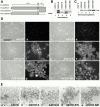
Figure 1
Figure 1
Expression of E/N- and N/E-cadherin in A431D cells. (A) Chimeric cadherins consisting of the extracellular and transmembrane domains of E-cadherin (white) and the cytoplasmic domain of N-cadherin (gray) or consisting of the extracellular and transmembrane domains of N-cadherin and the cytoplasmic domain of E-cadherin were cloned into pLKneo2. (B) A431D cells were transfected with N-cadherin, E-cadherin, E/N-cadherin, or N/E-cadherin and examined for transgene expression by immunoblotting with antibodies against the cytoplasmic domain of N-cadherin (lanes 1, 2, and 4), the extracellular domain of N-cadherin (lane 5), or the extracellular domain of E-cadherin (lane 3). Note, in some cases, we observed various processing variants when transfected cadherins were overexpressed in cells. (C) Extracts were immunoprecipitated, resolved by SDS-PAGE, and immunoblotted for β-catenin. (D) Untransfected A431D cells (a–d) or A431D cells expressing N-cadherin (e and f), E-cadherin (g and h), E/N-cadherin (f and g), or N/E-cadherin (h and i) were processed for immunofluorescence microscopy using the appropriate cadherin antibody. Corresponding phase and fluorescence micrographs are shown. (E) Untransfected A431 D cells (a) or A431D cells expressing E-cadherin (b), N-cadherin (c), E/N-cadherin (d), or N/E-cadherin (e) were tested for their ability to aggregate in a hanging drop aggregation assay. Bar, 10 μm.
Figure 4
Figure 4
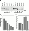
Generation of additional cadherin chimeras. (A) Chimeric cadherins, with a 2X-myc tag at the COOH terminus, consisting of E-cadherin (white) and N-cadherin (gray), were cloned into pLKpac. (B) A431D cells were transfected and examined for transgene expression by immunoblotting with anti-myc. Note, in some cases, we observed various processing variants when transfected cadherins were overexpressed in cells. (C) Extracts were immunoprecipitated using anti-myc, resolved by SDS-PAGE, and immunoblotted for β-catenin. (D) A431D cells expressing N/E-myc-cadherin (a), N/E5-myc-cadherin (b), N/E4-myc-cadherin (c), or N/E3-myc-cadherin (d) were tested for their ability to aggregate in a hanging drop aggregation assay. Bar, 15 μm.
Figure 4
Figure 4
Generation of additional cadherin chimeras. (A) Chimeric cadherins, with a 2X-myc tag at the COOH terminus, consisting of E-cadherin (white) and N-cadherin (gray), were cloned into pLKpac. (B) A431D cells were transfected and examined for transgene expression by immunoblotting with anti-myc. Note, in some cases, we observed various processing variants when transfected cadherins were overexpressed in cells. (C) Extracts were immunoprecipitated using anti-myc, resolved by SDS-PAGE, and immunoblotted for β-catenin. (D) A431D cells expressing N/E-myc-cadherin (a), N/E5-myc-cadherin (b), N/E4-myc-cadherin (c), or N/E3-myc-cadherin (d) were tested for their ability to aggregate in a hanging drop aggregation assay. Bar, 15 μm.
Figure 2
Figure 2
Expression of E/N- and N/E-cadherin in SCC1 cells. (A) SCC1 cells were transfected with E/N- or N/E-cadherin and examined for transgene expression by immunoblotting with antibodies against the cytoplasmic (lane 1) or extracellular (lane 2) domain of N-cadherin. (B) Extracts were immunoprecipitated, resolved by SDS-PAGE, and immunoblotted for β-catenin. (C) Untransfected SCC1 cells (a and b) or SCC1 cells expressing N-cadherin (c and d), E/N-cadherin (e and f), or N/E-cadherin (g and h) were processed for immunofluorescence microscopy using the appropriate cadherin antibody. Corresponding phase and fluorescence micrographs are shown. Bar, 15 μm.
Figure 2
Figure 2
Expression of E/N- and N/E-cadherin in SCC1 cells. (A) SCC1 cells were transfected with E/N- or N/E-cadherin and examined for transgene expression by immunoblotting with antibodies against the cytoplasmic (lane 1) or extracellular (lane 2) domain of N-cadherin. (B) Extracts were immunoprecipitated, resolved by SDS-PAGE, and immunoblotted for β-catenin. (C) Untransfected SCC1 cells (a and b) or SCC1 cells expressing N-cadherin (c and d), E/N-cadherin (e and f), or N/E-cadherin (g and h) were processed for immunofluorescence microscopy using the appropriate cadherin antibody. Corresponding phase and fluorescence micrographs are shown. Bar, 15 μm.
Figure 3
Figure 3
Expression of E/N- and N/E-cadherin in BT20 cells. (A) BT20 cells were transfected with full-length N-cadherin (BT20N), E/N-cadherin, or N/E-cadherin. Untransfected BT20 cells (a and b) or BT20 cells expressing N-cadherin (c and d), E/N-cadherin (e and f), or N/E-cadherin (g and h) were processed for immunofluorescence microscopy using the appropriate cadherin antibody. Corresponding phase and fluorescence micrographs are shown. Bar, 15 μm. (B) Cells were plated on membranes for motility assays, incubated for 24 h, and the number traversing the membrane was determined by averaging 10 random fields. Data are expressed as the number of cells/field. Each experiment was done three times and error bars indicate SD.
Figure 3
Figure 3
Expression of E/N- and N/E-cadherin in BT20 cells. (A) BT20 cells were transfected with full-length N-cadherin (BT20N), E/N-cadherin, or N/E-cadherin. Untransfected BT20 cells (a and b) or BT20 cells expressing N-cadherin (c and d), E/N-cadherin (e and f), or N/E-cadherin (g and h) were processed for immunofluorescence microscopy using the appropriate cadherin antibody. Corresponding phase and fluorescence micrographs are shown. Bar, 15 μm. (B) Cells were plated on membranes for motility assays, incubated for 24 h, and the number traversing the membrane was determined by averaging 10 random fields. Data are expressed as the number of cells/field. Each experiment was done three times and error bars indicate SD.
Figure 5
Figure 5
Expression of additional N/E-cadherin chimeras. (A) SCC1 cells were transfected with N/E-myc-cadherin, N/E5-myc-cadherin, N/E4-myc-cadherin, or N/E3-myc-cadherin and examined for transgene expression by immunoblotting with anti-myc. (B) Extracts were immunoprecipitated using anti-myc, resolved by SDS-PAGE, and immunoblotted for β-catenin. (C) SCC1 cells transfected with N/E-myc-cadherin (a and b) N/E5-myc-cadherin (c and d), N/E4-myc-cadherin (e and f), or N/E3-myc-cadherin (g and h) were processed for immunofluorescence microscopy using anti-myc. Corresponding phase and fluorescence micrographs are shown. Bar, 15 μm. (D) A431D cells either nontransfected or transfected with N-cadherin (A431D-N), E-cadherin (A431D-E), E/N-myc-cadherin (A431D-E/N), N/E-myc-cadherin (A431D-N/E), N/E5-myc-cadherin (A431D-N/E5), N/E4-myc-cadherin (A431D-N/E4), or N/E3-myc-cadherin (A431D-N/E3) were plated on membranes for motility assays, incubated for 24 h, and the number traversing the membrane was determined by averaging 10 random fields. Data are expressed as the number of cells/field. Each experiment was done three times and error bars indicate SD.
Figure 5
Figure 5
Expression of additional N/E-cadherin chimeras. (A) SCC1 cells were transfected with N/E-myc-cadherin, N/E5-myc-cadherin, N/E4-myc-cadherin, or N/E3-myc-cadherin and examined for transgene expression by immunoblotting with anti-myc. (B) Extracts were immunoprecipitated using anti-myc, resolved by SDS-PAGE, and immunoblotted for β-catenin. (C) SCC1 cells transfected with N/E-myc-cadherin (a and b) N/E5-myc-cadherin (c and d), N/E4-myc-cadherin (e and f), or N/E3-myc-cadherin (g and h) were processed for immunofluorescence microscopy using anti-myc. Corresponding phase and fluorescence micrographs are shown. Bar, 15 μm. (D) A431D cells either nontransfected or transfected with N-cadherin (A431D-N), E-cadherin (A431D-E), E/N-myc-cadherin (A431D-E/N), N/E-myc-cadherin (A431D-N/E), N/E5-myc-cadherin (A431D-N/E5), N/E4-myc-cadherin (A431D-N/E4), or N/E3-myc-cadherin (A431D-N/E3) were plated on membranes for motility assays, incubated for 24 h, and the number traversing the membrane was determined by averaging 10 random fields. Data are expressed as the number of cells/field. Each experiment was done three times and error bars indicate SD.
Figure 6
Figure 6
Generation and expression of E/N/E- and N/E/N-cadherins. (A) Chimeric cadherins consisting of E-cadherin (white) and N-cadherin (gray) were constructed and cloned into pLKpac with a 2X-myc tag at the COOH terminus. (B) A431D cells were transfected and examined for transgene expression by immunoblotting with anti-myc (lanes 1 and 2). Extracts were immunoprecipitated using anti-myc, resolved by SDS-PAGE, and immunoblotted for β-catenin (lanes 3 and 4). (C) A431D cells expressing E/N/E-myc-cadherin (a) or N/E/N-myc-cadherin (b) were tested for their ability to aggregate in a hanging drop aggregation assay. (D) SCC1 cells transfected with E/N/E-myc-cadherin (a and b) or N/E/N-myc-cadherin (c and d) were processed for immunofluorescence microscopy using anti-myc. Corresponding phase and fluorescence micrographs are shown. Bar, 15 μm. (E) A431D cells transfected with E-cadherin (A431D-E), N-cadherin (A431D-N), E/N/E-myc-cadherin (E/N/E), or N/E/N-myc-cadherin (N/E/N) were plated on membranes for motility assays, incubated for 24 h, and the number traversing the membrane was determined by averaging 10 random fields. Data are expressed as the number of cells/field. Each experiment was done three times and error bars indicate SD.
Figure 6
Figure 6
Generation and expression of E/N/E- and N/E/N-cadherins. (A) Chimeric cadherins consisting of E-cadherin (white) and N-cadherin (gray) were constructed and cloned into pLKpac with a 2X-myc tag at the COOH terminus. (B) A431D cells were transfected and examined for transgene expression by immunoblotting with anti-myc (lanes 1 and 2). Extracts were immunoprecipitated using anti-myc, resolved by SDS-PAGE, and immunoblotted for β-catenin (lanes 3 and 4). (C) A431D cells expressing E/N/E-myc-cadherin (a) or N/E/N-myc-cadherin (b) were tested for their ability to aggregate in a hanging drop aggregation assay. (D) SCC1 cells transfected with E/N/E-myc-cadherin (a and b) or N/E/N-myc-cadherin (c and d) were processed for immunofluorescence microscopy using anti-myc. Corresponding phase and fluorescence micrographs are shown. Bar, 15 μm. (E) A431D cells transfected with E-cadherin (A431D-E), N-cadherin (A431D-N), E/N/E-myc-cadherin (E/N/E), or N/E/N-myc-cadherin (N/E/N) were plated on membranes for motility assays, incubated for 24 h, and the number traversing the membrane was determined by averaging 10 random fields. Data are expressed as the number of cells/field. Each experiment was done three times and error bars indicate SD.
Figure 7
Figure 7
Anti–N-cadherin mAb 8C11 inhibits motility. (A) Extracts of A431D cells transfected with N/E-myc-cadherin (lanes 1 and 7), N/E5a-myc-cadherin (lanes 2 and 8), N/E5-myc-cadherin (lanes 3 and 9), N/E4-myc-cadherin (lanes 4 and 10), N/E3-myc-cadherin (lanes 5 and 11), or N/E2-myc-cadherin (lanes 6 and 12) were resolved by SDS-PAGE and immunoblotted with mAb 8C11 (lanes 1–6) or anti-myc (lanes 7–12). Note, in some cases, we observed various processing variants when transfected cadherins were overexpressed in cells. (B) BT20 cells, which were transfected with N-cadherin (BT20N), were plated on membranes for motility assays in the presence of no antibody or 8C11 ascites at a dilution of 1:10–1:100. Untransfected BT20 cells in the absence of antibody were included as a control. After 24 h, the number of cells traversing the membrane was determined by averaging 10 random fields at 100× magnification. Data are expressed as the number of cells/field. (C) Untransfected BT20 cells or BT20 cells transfected with N-cadherin (BT20N) were plated on membranes for motility assays in the presence of no antibody, irrelevant ascites 4A2 at a dilution of 1:10 or 8C11 ascites at a dilution of 1:10. After 24 h, the number of cells traversing the membrane was determined by counting the entire membrane. Data are expressed as the number of cells traversing the filter.

References

    1. Aono S., Nakagawa S., Reynolds A.B., Takeichi M. p120(ctn) acts as an inhibitory regulator of cadherin function in colon carcinoma cells. J. Cell Biol. 1999;145:551–562. - PMC - PubMed
    1. Behrens J. Cadherins and cateninsrole in signal transduction and tumor progression. Cancer Metastasis Rev. 1999;18:15–30. - PubMed
    1. Blaschuk O.W., Sullivan R., David S., Pouliot Y. Identification of a cadherin cell adhesion recognition sequence. Dev. Biol. 1990;139:227–229. - PubMed
    1. Brieher W.M., Yap A.S., Gumbiner B.M. Lateral dimerization is required for the homophilic binding activity of C-cadherin. J. Cell Biol. 1996;135:487–496. - PMC - PubMed
    1. Byers S., Amaya E., Munro S., Blaschuk O. Fibroblast growth factor receptors contain a conserved HAV region common to cadherins and influenza strain A hemagglutininsa role in protein–protein interactions? Dev. Biol. 1992;152:411–414. - PubMed

Publication types