. 2000 Dec 11;151(6):1345-52.
doi: 10.1083/jcb.151.6.1345.

Numb is an endocytic protein

Affiliations

Numb is an endocytic protein

E Santolini et al. J Cell Biol. .

Abstract

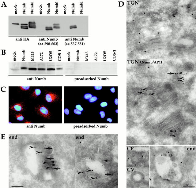
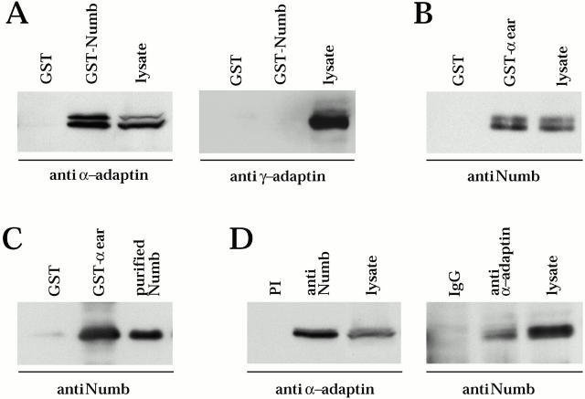
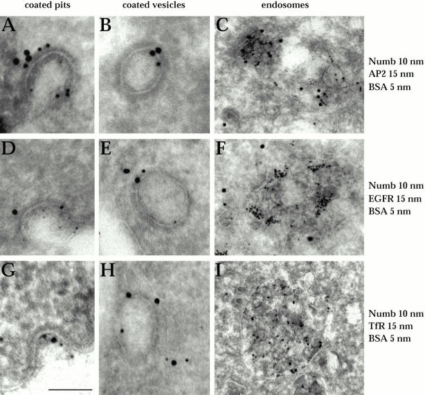
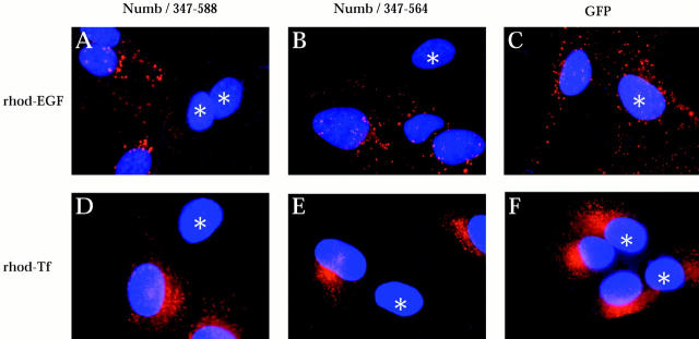
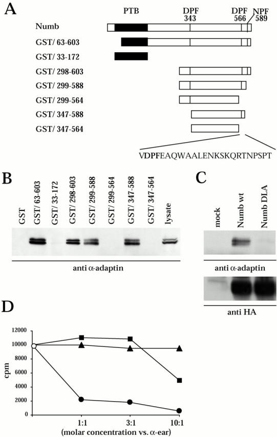
Numb is a protein that in Drosophila determines cell fate as a result of its asymmetric partitioning at mitosis. The function of Numb has been linked to its ability to bind and to biologically antagonize Notch, a membrane receptor that also specifies cell fate. The biochemical mechanisms underlying the action of Numb, however, are still largely unknown. The wide pattern of expression of Numb suggests a general function in cellular homeostasis that could be additional to, or part of, its action in fate determination. Such a function could be endocytosis, as suggested by the interaction of Numb with Eps15, a component of the endocytic machinery. Here, we demonstrate that Numb is an endocytic protein. We found that Numb localizes to endocytic organelles and is cotrafficked with internalizing receptors. Moreover, it associates with the appendage domain of alpha adaptin, a subunit of AP2, a major component of clathrin-coated pits. Finally, fragments of Numb act as dominant negatives on both constitutive and ligand-regulated receptor-mediated internalization, suggesting a general role for Numb in the endocytic process.


Figures

Figure 1
Figure 1
Numb localizes to endocytic organelles. (A) HeLa cells transfected with HA-Numb (Numb), HA-Numbl (Numbl), or mock transfected (mock) were analyzed by immunoblotting with the antibodies indicated underneath. (B) Cellular lysates from the indicated cell lines, as well as from the mock- and HA-Numb–transfected HeLa were analyzed with the anti-Numb antibody (aa 537–551) or with the same antibody preabsorbed with the cognate peptide. (C) CV1 cells were stained with the anti-Numb antibody (aa 537–551) or with same antibody preabsorbed with the cognate peptide, as indicated. Similar results were observed in HeLa, COS-1, A172, M413, U2OS, and VA13 cells (data not shown). (D) Immunoelectron microscopy performed on exponentially growing A172 cells. In all panels, except the TGN (Numb/AP1) panel, staining was with the anti-Numb antibody (aa 537–551, 15 nm gold). BSA was used as a tracer of endocytosis by incubating the cells for 1 h with BSA-gold/5 nm before fixation. Representative examples are shown of the TGN, coated pits (CP), coated vesicles (CV), and endosomes (end). In the TGN (Numb/AP1) panel, a double labeling for Numb and γ adaptin was performed (Numb, 15 nm gold, arrows; γ adaptin, 10 nm gold, arrowheads). (e) Immunoelectron microscopy performed on A172 cells with the anti-Numb (aa 537–551, 10 nm gold, arrows) and anti-Eps15 antibodies (15 nm gold, arrowheads). Two representative endosomes are shown. Bars: (E and D, TGN) 0.18 μm; (D, TGN Numb/AP1) 0.16 μm; (D, CP) 0.22 μm; (D, CV) 0.25 μm; (D, end) 0.20 μm.
Figure 3
Figure 3
Interaction between Numb and α adaptin. (a) GST-Numb/63–603 (GST-Numb) or control GST was incubated with COS-1 cell lysates followed by immunoblotting with the indicated antibodies. In this and all subsequent panels, the lanes referred to as lysate were loaded with 100 μg of the cellular lysates to serve as a reference. (b) A GST fusion protein containing aa 715–977 of the mouse α adaptin (GST–α ear) or GST alone was incubated with a cellular lysate from A172 cells and immunoblotted with the anti-Numb antibody (aa 537–551). (c) GST–α ear was incubated with a fragment of Numb (aa 298–603), expressed in bacteria as a GST fusion protein, thrombin cleaved, and purified. An aliquot of the purified Numb fragment (purified Numb) was also loaded to serve as a reference. (d) A172 cellular lysates were immunoprecipitated with either the anti-Numb antibody (aa 298–603) or with the corresponding preimmune serum and immunoblotted with anti–α adaptin (left). A172 cell lysate were also immunoprecipitated with an anti–α adaptin mAb or with mouse IgG as a control, followed by detection with anti-Numb antibody (aa 537–551) (right).
Figure 2
Figure 2
Numb cotrafficks with internalizing receptors. A172 cells were serum starved for 16–20 h followed by incubation with 100 ng/ml EGF and BSA-gold/5 nm for 1 h on ice, and temperature-shifted at 37°C for 2 min (A and B, D and E, G and H) or 10 min (C, F, and I). (A–C) Colocalization of Numb (10 nm gold) with AP2 (15 nm gold). (d–f) Colocalization of Numb (10 nm gold) with EGFR (15 nm gold). (g–i) Colocalization of Numb (10 nm gold) with TfR (15 nm gold). Representative examples are shown: A, D and g, coated pits; b, e, and h, coated vesicles; c, f, and i, endosomes. Bar: (a and b) 0.10 μm; (c) 0.17 μm; (d, e, and g) 0.13 μm; (f and h) 0.15 μm; (i) 0.22 μm.
Figure 5
Figure 5
Numb inhibits clathrin-mediated internalization. CV1 cells were transfected with pCDNA3 expression vectors encoding either HA-Numb/347–588 (a and d) or HA-Numb/347–564 (b and e), or GFP as a control (c and f). The cells were then incubated with either rhodamine-conjugated EGF (a–c) or rhodamine-conjugated Tf (d–f). Transfected cells were detected with either an anti-Numb antibody (a, b, d, and e) or by direct GFP fluorescence (c and f), and are marked with asterisks. Internalized EGF or Tf is visible in red. More than 50% of the cells transfected with Numb deletion mutants presented a complete block in EGF and Tf uptake in three independent experiments. Transfection of full-length Numb did not alter the initial rate of endocytosis of both EGFR and TfR (data not shown).
Figure 4
Figure 4
Mapping of the Numb–AP2 interaction. (a) Schematic of the GST fusion proteins employed. The PTB domain is shown in black. The positions of the DPF and NPF motifs are also indicated with vertical bars and aa positions. The aa sequence of positions 564–588 is also shown. (b) The fusion proteins, depicted in A, were incubated with cellular lysates from COS-1 cells, followed by immunoblotting with an anti–α adaptin antibody. (c) The DPF motif in position 566–568 of Numb was mutagenized to DLA, and the HA-tagged versions of the wild-type and mutated Numb cDNAs were transfected in HeLa cells and immunoprecipitated with the anti-HA antibody, followed by immunoblotting with anti–α adaptin (top) or with anti-HA (bottom). (d) Competition between Numb and Eps15 for binding to α ear (○, no competition; ▪, competition with Numb (aa 298-603); •, competition with Eps15 (aa 581-874); ▴, control competition with BSA. For details see Materials and Methods.

References

    1. Artavanis-Tsakonas S., Matsuno K., Fortini M.E. Notch signaling. Science. 1995;268:225–232. - PubMed
    1. Artavanis-Tsakonas S., Rand M.D., Lake R.J. Notch signalingcell fate control and signal integration in development. Science. 1999;284:770–776. - PubMed
    1. Benmerah A., Gagnon J., Begue B., Megarbane B., Dautry-Varsat A., Cerf-Bensussan N. The tyrosine kinase substrate eps15 is constitutively associated with the plasma membrane adaptor AP-2. J. Cell Biol. 1995;131:1831–1838. - PMC - PubMed
    1. Benmerah A., Begue B., Dautry-Varsat A., Cerf-Bensussan N. The ear of alpha-adaptin interacts with the COOH-terminal domain of the Eps 15 protein. J. Biol. Chem. 1996;271:12111–12116. - PubMed
    1. Berezovska O., McLean P., Knowles R., Frosh M., Lu F.M., Lux S.E., Hyman B.T. Notch1 inhibits neurite outgrowth in postmitotic primary neurons. Neuroscience. 1999;93:433–439. - PubMed

Publication types

MeSH terms