. 2000 Dec 15;28(24):4832-8.
doi: 10.1093/nar/28.24.4832.

Interaction of human DNA topoisomerase I with G-quartet structures

Affiliations

Interaction of human DNA topoisomerase I with G-quartet structures

P B Arimondo et al. Nucleic Acids Res. .

Abstract

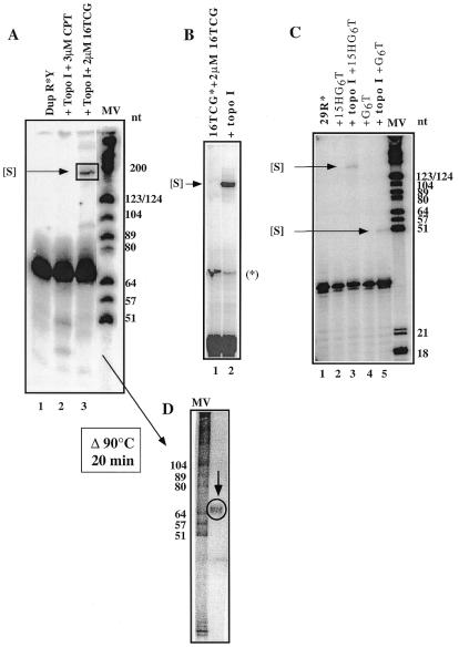
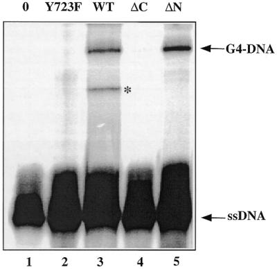
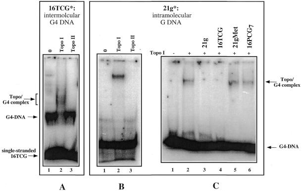
Because of their role in the control of the topological state of DNA, topoisomerases are ubiquitous and vital enzymes, which participate in nearly all events related to DNA metabolism including replication and transcription. We show here that human topoisomerase I (Topo I) plays an unexpected role of 'molecular matchmaker' for G-quartet formation. G-quadruplexes are multi-stranded structures held together by square planes of four guanines ('G-quartets') interacting by forming Hoogsteen hydrogen bonds. Topo I is able to promote the formation of four-stranded intermolecular DNA structures when added to single-stranded DNA containing a stretch of at least five guanines. We provide evidence that these complexes are parallel G-quartet structures, mediated by tetrads of hydrogen-bonded guanine. In addition, Topo I binds specifically to pre-formed parallel and anti-parallel G4-DNA.


Figures

Figure 1
Figure 1
Formation of a complex of slower mobility [S]. A weight marker was used (molecular weight MV, Boehringer Mannheim). The oligopurine-containing strand of the duplex is indicated by R and the oligopyrimidine sequence by Y. Oligonucleotide sequences are given in Table 1. (A) The 69 bp duplex was 3′ end-radiolabelled on the purine strand (lane 1) and was incubated for 1 h at 20°C as described in the presence of 3 µM CPT (lane 2) or in the presence of 2 µM 16TCG (lane 3). The samples were analysed on a denaturing 15% polyacrylamide gel. (B) 20 nM of 3′ end-radiolabelled 16TCG was incubated with 2 µM of unlabelled 16TCG oligonucleotide (lane 1) and after addition of Topo I (lane 2) and proteolyses, the samples were analysed on a sequencing gel. The band marked with an asterisk corresponds to a labelling artefact (radiolabelled dimer). (C) 20 nM of 3′ end-radiolabelled 29R was incubated with 2 µM of 15HG6T oligonucleotide (lanes 2 and 3) or 2 µM of G6T oligonucleotide (lanes 4 and 5) in the absence (lanes 2 and 4) or in the presence of Topo I (lanes 3 and 5). (D) The complex of slower mobility was cut, purified and heated for 20 min at 90°C prior to reloading on a sequencing gel.
Figure 2
Figure 2
The complex is four-stranded. (A) Model structure of a guanine quartet and a four-stranded intermolecular complex. (B) Determination of the stoichiometry of the complex induced by Topo I between radiolabelled oligonucleotide 29R (A) and oligonucleotide G6T (B). The tetrameric species are indicated on the right. The arrows indicate the free probe and the G4–DNA structures.
Figure 3
Figure 3
Role of Topo I. The 69 bp duplex after preincubation with 2 µM 16TCG in the described buffer was treated with different Topo I mutants: lane 1, no enzyme; lane 2, Topo I mutant Y723F; lane 3, Topo I wild-type; lane 4, Topo I mutant ΔC; lane 5, Topo I mutant ΔN. After protease digestion, the samples were loaded on a sequencing gel. The presence of the band marked with an asterisk was dependent on the Topo I preparation in the experiment and was not further characterised.
Figure 4
Figure 4
Topo I binds to intermolecular and intramolecular G4–DNA. 20 nM of radiolabelled 16TCG and 2 µM of non-radiolabelled 16TCG preincubated in KCl (A) or 20 nM of radiolabelled 21g (B) were incubated for 15min at 20°C in the absence (lane 1) or in the presence of 0.4 nM Topo I (lane 2) or Topo II (lane 3). The samples were analysed by gel electrophoresis in 0.25× TBE onto a 6% polyacrylamide gel [29:1 acrylamide:bisacrylamide]. The arrows indicate the free probe, the G4–DNA structure and the Topo I/G4–DNA complex (Topo/G4 complex). (C) Competition experiments. 20 nM of radiolabelled 21g were incubated in the above buffer (lane 1) and in the presence of Topo I (lane 2) with a 1000-fold excess of: 21g (lane 3), 16TCG (lane 4), 21gMet (lane 5) and 16PCG7 (lane 6). The 21gMet oligonucleotide was obtained by DMS treatment for 15 min at 37°C from the 21g oligonucleotide; after this treatment the oligonucleotide does not form G-quartets structures. Only the quadruplex-forming oligonucleotides (lanes 3 and 4) are therefore able to compete. The oligonucleotides sequences are reported in Table 1.

References

    1. Wang J.C. (1996) DNA topoisomerases. Annu. Rev. Biochem., 65, 635–692. - PubMed
    1. Nitiss J.L. (1998) Investigating the biological functions of DNA topoisomerases in eukaryotic cells. Biochim. Biophys. Acta, 1400, 63–81. - PubMed
    1. Pommier Y., Pourquier,P., Fan,Y. and Strumberg,D. (1998) Mechanism of action of eukaryotic DNA topoisomerase I and drugs targeted to the enzyme. Biochim. Biophys. Acta, 1400, 83–105. - PubMed
    1. Redinbo M.R., Stewart,L., Kuhn,P., Champoux,J.J. and Hol,W.G. (1998) Crystal structures of human topoisomerase I in covalent and noncovalent complexes with DNA. Science, 279, 1504–1513. - PubMed
    1. Sen D. and Gilbert,W. (1988) Formation of parallel four-stranded complexes by guanine-rich motifs in DNA and its applications for meiosis. Nature, 334, 364–366. - PubMed

Publication types

MeSH terms