. 2000 Dec 15;28(24):4998-5004.
doi: 10.1093/nar/28.24.4998.

Quantitative analysis of globin gene induction in single human erythroleukemic cells

Affiliations

Quantitative analysis of globin gene induction in single human erythroleukemic cells

R D Smith et al. Nucleic Acids Res. .

Abstract

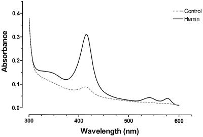
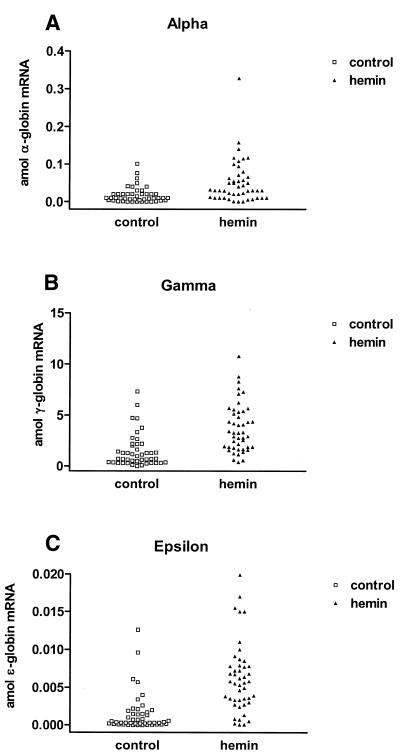
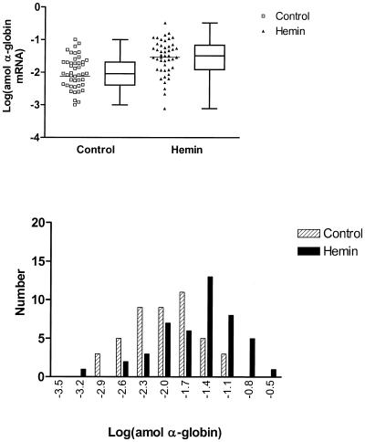
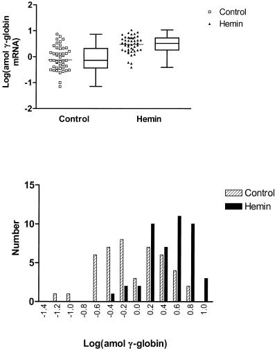
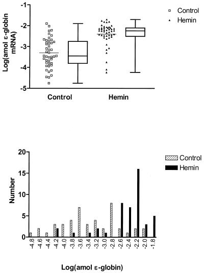
The mechanisms involved in the normal developmental regulation of globin gene expression, and the response to pharmacological agents that elevate fetal hemoglobin, may be expected to involve either changes in each cell or a selection process affecting subsets of differentiating erythroid cells. To study these mechanisms we have developed assays to measure mRNA levels in single erythroid cells. The assay involved the use of globin-specific probes, with no detectable cross-reactivity, in real-time, fluorescence-based quantitative PCR (Q-PCR). We had previously used this Q-PCR method to measure globin mRNA levels in cultures of primary erythroid cells demonstrating that drugs like hydroxyurea, 5-azacytidine and butyric acid each yielded increases in gamma/( gamma + ss) mRNA ratios, with differential effects on ss-globin levels. We have now extended this approach to measure globin mRNA levels in single K562 cells, a human erythroleukemic cell line, with and without 30 microM hemin treatment. Hemin exposure increases total hemoglobin levels by approximately 9-fold and total alpha-, epsilon- and gamma-globin mRNA levels by 1.5-2.3-fold. Single cell analyses showed initial wide distributions of each of the three individual globin mRNA levels with most cells having detectable but very low levels of each globin transcript. Hemin induction shifted the distributions to higher levels, with a tendency to residual left skewing as some cells remained with very low expression levels despite the effect of hemin in increasing expression in most of these low expressing cells. Thus transcriptional heterogeneity remains a crucial variable, even in this extensively used model of human erythroid biology, and clearly influences strongly the response to inducing agents. These methods may enable us to define better possible molecular and/or cellular models of globin gene modulation.


Figures

Figure 1
Figure 1
Absorption spectroscopy of K562 cell lysates. Spectra of lysates from control (dashed line) and cells treated with 30 µM hemin for 48 h (solid line) are shown from 300 to 625 nm. The change in absorbance at 413 nm normalized to the protein concentration (ΔA413/mg total protein) was determined and used as an indicator of hemoglobin expression level.
Figure 2
Figure 2
Plots of transcript levels for control (squares) and 30 µM hemin treated (triangles) K562 cells. Data for α-globin (A), γ-globin (B) and ɛ-globin (C) are shown. Mean values for globin transcript levels were as follows: 1.4 ± 1.6 amol/cell γ-globin, 0.017 ± 0.021 amol/cell α-globin and 0.0015 ± 0.0025 amol/cell ɛ-globin for control cells increasing to 3.7 ± 2.4 amol/cell γ-globin, 0.052 ± 0.058 amol/cell α-globin and 0.0062 ± 0.0046 amol/cell ɛ-globin respectively for hemin-treated cells (mean ± standard deviation).
Figure 3
Figure 3
Logarithmic transformation of α-globin transcript levels in single K562 cells. The upper panel presents plots of transformed data for single-cell expression in control (squares) and hemin-treated (triangles) K562 cells. The bars represent the mean values for logarithm of the globin level which increased from –2.13 to –1.54 (P = 0.0004, t-test), respectively. Concomitantly, the variance declined from 0.90 to 0.28 (P < 0.0001, F-test). Box and whisker plots are presented alongside the scatter plots showing the 25th and 75th percentile and median values for transcript level. Frequency histograms of the transformed data are shown in the lower panel with controls shown as hatched columns and hemin-treated cells as solid columns. The skew in the distributions for the transformed data for control and treated cells is 0.001 (P = 1.00) and –0.690 (P = 0.041), respectively.
Figure 4
Figure 4
Logarithmic transformation of γ-globin transcript levels in single K562 cells. The upper panel presents plots of transformed data for single cell expression in control (squares) and hemin-treated (triangles) K562 cells in which the mean (bars) was increased from –0.12 to 0.47 (P < 0.0001, t-test) respectively while the variance was decreased from 0.40 to 0.10 (P < 0.0001, F-test). Box and whisker plots are presented alongside the scatter plots showing the 25th and 75th percentile and median values for transcript level. Frequency histograms of the transformed data are shown in the lower panel with controls shown as hatched columns and hemin-treated cells as solid columns. The skew in the distributions for the transformed data for control and treated cells is –0.025 (P = 0.94) and –0.658 (P = 0.052) respectively.
Figure 5
Figure 5
Logarithmic transformation of ɛ-globin transcript levels in single K562 cells. The upper panel presents plots of transformed data for single-cell expression in control (squares) and hemin-treated (triangles) K562 cells in which the mean (bars) was increased from –3.30 to –2.40 (P < 0.0001, t-test), respectively, while the variance was decreased from 0.49 to 0.31 (P = 0.06, F-test). Box and whisker plots are presented alongside the scatter plots showing the 25th and 75th percentile and median values for transcript level. Frequency histograms of the transformed data are shown in the lower panel with controls shown as hatched columns and hemin-treated cells as solid columns. The skew in the distributions for the transformed data for control and treated cells is 0.005 (P = 0.988) and –1.781 (P < 0.001) respectively.

Similar articles

Cited by

References

    1. Baron M.H. (1996) Developmental regulation of the vertebrate globin multigene family. Gene Expr., 6, 129–137. - PMC - PubMed
    1. Rodgers G.P. and Rachmilewitz,E.A. (1995) Novel treatment options in the severe β-globin disorders. Br. J. Haematol., 91, 263–268. - PubMed
    1. Olivieri N.F. and Weatherall,D.J. (1998) The therapeutic reactivation of fetal haemoglobin. Hum. Mol. Genet., 7, 1655–1658. - PubMed
    1. Atweh G.F., Sutton,M., Nassif,I., Boosalis,V., Dover,G.J., Wallenstein,S., Wright,E., McMahon,L., Stamatoyannopoulos,G., Faller,D.V. and Perrine,S.P. (1999) Sustained induction of fetal hemoglobin by pulse butyrate therapy in sickle cell disease. Blood, 93, 1790–1797. - PMC - PubMed
    1. Snabes M.C., Chong,S.S., Subramanian,S.B., Kristjansson,K., DiSepio,D. and Hughes,M.R. (1994) Preimplantation single-cell analysis of multiple genetic loci by whole-genome amplification. Proc. Natl Acad. Sci. USA, 91, 6181–6185. - PMC - PubMed

MeSH terms