. 2000 Dec 15;14(24):3115-25.
doi: 10.1101/gad.851100.

Structural basis of inhibition of CDK-cyclin complexes by INK4 inhibitors

Affiliations

Structural basis of inhibition of CDK-cyclin complexes by INK4 inhibitors

P D Jeffrey et al. Genes Dev. .

Abstract

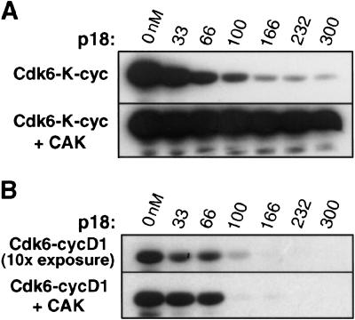
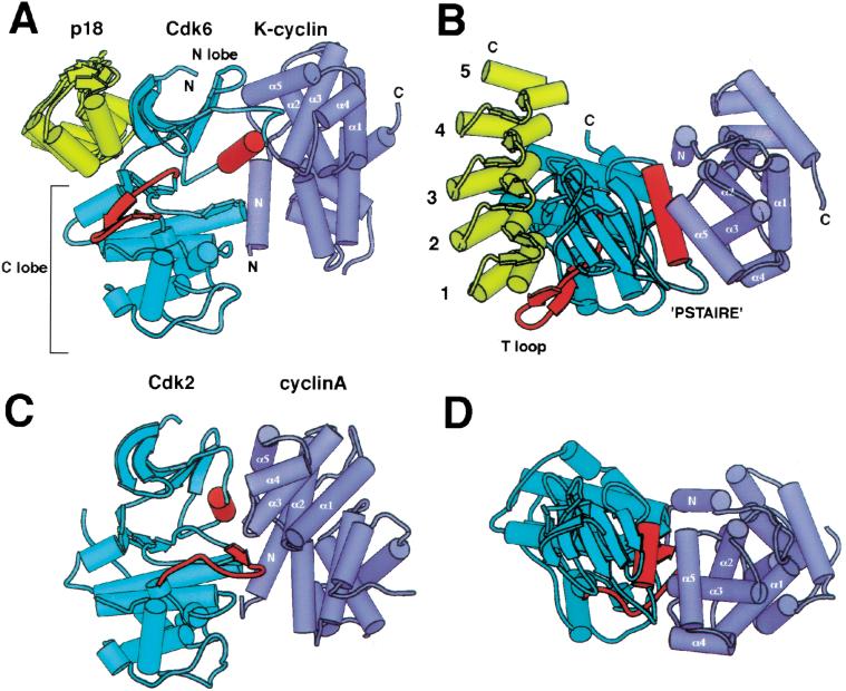
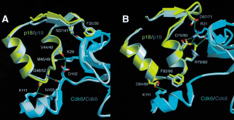
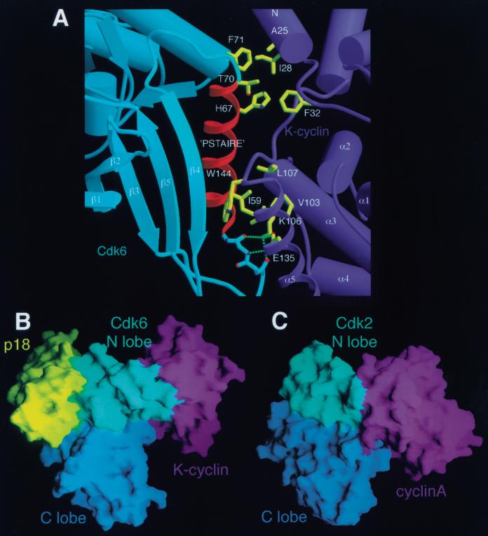
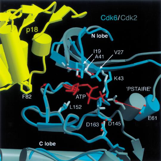
The cyclin-dependent kinases 4 and 6 (Cdk4/6) that drive progression through the G(1) phase of the cell cycle play a central role in the control of cell proliferation, and CDK deregulation is a frequent event in cancer. Cdk4/6 are regulated by the D-type cyclins, which bind to CDKs and activate the kinase, and by the INK4 family of inhibitors. INK4 proteins can bind both monomeric CDK, preventing its association with a cyclin, and also the CDK-cyclin complex, forming an inactive ternary complex. In vivo, binary INK4-Cdk4/6 complexes are more abundant than ternary INK4-Cdk4/6-cyclinD complexes, and it has been suggested that INK4 binding may lead to the eventual dissociation of the cyclin. Here we present the 2.9-A crystal structure of the inactive ternary complex between Cdk6, the INK4 inhibitor p18(INK4c), and a D-type viral cyclin. The structure reveals that p18(INK4c) inhibits the CDK-cyclin complex by distorting the ATP binding site and misaligning catalytic residues. p18(INK4c) also distorts the cyclin-binding site, with the cyclin remaining bound at an interface that is substantially reduced in size. These observations support the model that INK4 binding weakens the cyclin's affinity for the CDK. This structure also provides insights into the specificity of the D-type cyclins for Cdk4/6.


Figures

Figure 1
Figure 1
The Rb kinase activity of the Cdk6–K-cyclin complex is inhibited by p18 when Cdk6 is unphosphorylated but not when Cdk6 is phosphorylated by the Cdk7–cyclinH CDK-activating kinase. (A) Phosphorylation of the Rb C-terminal fragment by the unphosphorylated Cdk6–K-cyclin complex (top panel) and the phosphorylated Cdk6–K-cyclin complex (lower panel). Both Cdk6 complexes are at 100 nM concentration. The lanes in the two panels contain p18 at 0, 33, 66, 100, 166, 232, and 300 nM concentrations. (B) Comparison with the Rb kinase activity of the Cdk6–cyclinD complex. Reactions contain the same concentration (100 nM) of Cdk6 and p18 as in the corresponding lanes in A. The gel in the bottom panel was exposed for the same length of time as the panels in A; the top panel showing the activity of unphosphorylated Cdk6–cyclinD complex was exposed 10-fold longer because of the low kinase activity of the unphosphorylated complex.
Figure 2
Figure 2
Overall structure of the p18–Cdk6–K-cyclin complex and comparison with Cdk2–cyclinA. (A) Schematic view of p18–Cdk6–K-cyclin. p18 is shown in yellow, Cdk6 in cyan, K-cyclin in purple. The T loop and PSTAIRE elements of Cdk6 are highlighted in red, and the helices of the first cyclin repeat are labeled. N and C termini are labeled where visible. The p18–Cdk6 and K-cyclin–Cdk6 interfaces do not overlap and lie on opposite sides of the kinase, burying a total of 4350 Å2 of surface area. (B) Top view of the p18–Cdk6–K-cyclin complex, approximately orthogonal to view in A. The ankyrin repeats of p18 are numbered. The PSTAIRE helix is central to the Cdk6–K-cyclin interface, but the T loop packs on the other side of the kinase. (C) View of Cdk2–cyclinA complex superimposed on the C lobe of Cdk6 in the same orientation as in A. Both the PSTAIRE helix and T loop, in red, pack against cyclinA. (D) View of superimposed Cdk2–cyclinA complex from same viewpoint as B.
Figure 3
Figure 3
Interactions between p18 and Cdk6 mirror those made between p19 and Cdk6. (A) Hydrogen bond networks between the second ankyrin repeat of p18 and Cdk6 involve equivalent residues to those in p19. p19–Cdk6 is superimposed on p18–Cdk6 using residues from the second ankyrin repeat and adjacent residues in the Cdk6 N lobe. The p18–Cdk6 residues are colored in yellow and cyan, that of p19–Cdk6 in gray. Hydrogen bonds in the p18–Cdk6 complex are indicated by green dashed lines. The same hydrogen bonds are made in the p19–Cdk6 complex but omitted here for clarity. Residue numbering for the INK4 inhibitors is given as p18/p19. (B) Interface between the third ankyrin repeat of p18 and Cdk6 showing the extensive hydrogen bonding network centered around Arg 31 of Cdk6. Residues from the second and third ankyrin repeats of p18 contribute most of the interactions with Cdk6.
Figure 4
Figure 4
The K-cyclin–Cdk6 interface. (A) The PSTAIRE helix of Cdk6 is a central feature of the Cdk6–K-cyclin interface. The viewpoint shown corresponds approximately to that in B. Three sets of interactions are shown: hydrogen bonds between the Cdk6 main-chain preceding the PSTAIRE helix and the conserved Lys–Glu pair of K-cyclin (K106, E135); the conserved Ile 59 of Cdk6 inserts into a hydrophobic pocket in K-cyclin; residues at the end of the PSTAIRE helix, one turn longer in Cdk4 and Cdk6 than in Cdk2, interact with residues on the N-terminal helix of K-cyclin and may play a role in cyclin–CDK specificity. (B) Surface representation of p18–Cdk6–K-cyclin complex illustrating the minimal interactions between K-cyclin and the Cdk6 C lobe. p18 is colored yellow, the Cdk6 N lobe is cyan, the Cdk6 C lobe is blue, and the K-cyclin is purple. The viewpoint is approximately the same as Fig. 2A. The only contacts between K-cyclin and the C lobe of Cdk6 arise from interactions with the N-terminal helix of K-cyclin. (C) Surface representation of Cdk2–cyclinA in the equivalent orientation as that in A, showing significantly greater interactions between the C lobe of the Cdk2 and the cyclinA, giving rise to a much more extensive cyclin–CDK interface.
Figure 5
Figure 5
Schematic representation of the different conformations of the CDK. CDKs undergo extensive conformational changes on binding of activating or inhibiting subunits. The major determinants of activity are the positions and conformation of the PSTAIRE helix and T loop, as well as the relative disposition of the kinase N and C lobes. The PSTAIRE helix adopts a position further away from the catalytic cleft in inactive CDKs (labeled as ‘out’) than in active CDKs (‘in’). The PSTAIRE helix conformation correlates with the location of a conserved active site residue (Cdk2, Glu 51; Cdk6, Glu 61) either inside or outside the catalytic cleft.
Figure 6
Figure 6
The ATP-binding site of p18–Cdkl6–K-cyclin and Cdk2–cyclinA. Active site residues implicated in ATP binding and catalysis are displaced in the p18–Cdk6–K-cyclin complex relative to the active Cdk2–cyclinA conformation. Cdk2 and Cdk6 were superimposed on their C lobes. Cdk6 is shown in cyan, p18 in yellow, Cdk2 in gray. Movement of active site residues is indicated by red arrows. p18 displaces the N lobe relative to the C lobe, causing the hydrophobic residues (Ile 19, Val 27, Ala 41, Leu 152) that sandwich the adenine ring of ATP to move by up to 4.5 Å. The p18 inhibitor also distorts the edge of the active site via Phe 82, affecting hydrogen bonding interactions with the edge of the ATP ring. The related shift of the PSTAIRE helix on the other side of the active site displaces an active site residue (Glu 61). The T loop of Cdk6 diverges from that of Cdk2 between Phe 164 and Val 181, and these residues are omitted for clarity.

References

    1. Adachi M, Roussel MF, Havenith K, Sherr CJ. Features of macrophage differentiation induced by p19INK4d, a specific inhibitor of cyclin D-dependent kinases. Blood. 1997;90:126–137. - PubMed
    1. Brotherton DH, Dhanaraj V, Wick S, Brizuela L, Domaille PJ, Volyanik E, Xu X, Parisini E, Smith BO, Archer SJ, et al. Crystal structure of the complex of the cyclin D-dependent kinase Cdk6 bound to the cell-cycle inhibitor p19INK4d. Nature. 1998;395:244–250. - PubMed
    1. Brown NR, Noble ME, Endicott JA, Johnson LN. The structural basis for specificity of substrate and recruitment peptides for cyclin-dependent kinases. Nat Cell Biol. 1999;1:438–443. - PubMed
    1. Brünger AT, Adams PD, Clore GM, Delano WL, Gros P, Grosse-Kunstieve RW, Jiang JS, Kuszewski J, Nilges M, Pannu NS. Crystallography and NMR system: A new software suite for macromolecular structure determination. Acta Crystallogr D. 1998;54:905–921. - PubMed
    1. Card GL, Knowles P, Laman H, Jones N, McDonald NQ. Crystal structure of a γ-herpesvirus cyclin–cdk complex. EMBO J. 2000;19:2877–2888. - PMC - PubMed

Publication types

MeSH terms

Associated data

LinkOut - more resources