. 2000 Dec 15;14(24):3126-39.
doi: 10.1101/gad.182800.

Mutation of a Drosophila gamma tubulin ring complex subunit encoded by discs degenerate-4 differentially disrupts centrosomal protein localization

Affiliations

Mutation of a Drosophila gamma tubulin ring complex subunit encoded by discs degenerate-4 differentially disrupts centrosomal protein localization

V Barbosa et al. Genes Dev. .

Abstract

We have cloned the Drosophila gene discs degenerate-4 (dd4) and find that it encodes a component of the gamma-tubulin ring complex (gammaTuRC) homologous to Spc98 of budding yeast. This provides the first opportunity to study decreased function of a member of the gamma-tubulin ring complex, other than gamma-tubulin itself, in a metazoan cell. gamma-tubulin is no longer at the centrosomes but is dispersed throughout dd4 cells and yet bipolar metaphase spindles do form, although these have a dramatically decreased density of microtubules. Centrosomin (CNN) remains in broad discrete bodies but only at the focused poles of such spindles, whereas Asp (abnormal spindle protein) is always present at the presumptive minus ends of microtubules, whether or not they are focused. This is consistent with the proposed role of Asp in coordinating the nucleation of mitotic microtubule organizing centers. The centrosome associated protein CP190 is partially lost from the spindle poles in dd4 cells supporting a weak interaction with gamma-tubulin, and the displaced protein accumulates in the vicinity of chromosomes. Electron microscopy indicates not only that the poles of dd4 cells have irregular amounts of pericentriolar material, but also that they can have abnormal centrioles. In six dd4 cells subjected to serial sectioning centrioles were missing from one of the two poles. This suggests that in addition to its role in nucleating cytoplasmic and spindle microtubules, the gammaTuRC is also essential to the structure of centrioles and the separation of centrosomes.


Figures
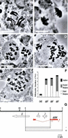
Figure 1
Figure 1
Mitotic defects revealed in orcein-stained preparations of the CNS of dd41 larvae. Panels A and B show respective metaphase and anaphase figures from wild-type female larvae. Panels C–E are from dd41mutant male larvae. Note the extreme chromatin condensation of mutant cells compared to the wild-type cells. Euploid (right) and 8N (left) metaphase cells (C). (D) An anaphase cell. We classify this cell as an anaphase as many sister chromatids appear to have separated and some appear as if pulled to poles. These cells are in fact rather difficult to classify, but resemble the catastrophic anaphases reported by Gatti and Baker (1989) for this mutant allele. (E) A 16N metaphase cell. (F) Proportions of euploid and polyploid metaphases and anaphase figures in three dd4 alleles in comparison to the wild-type strain Oregon-R. (G) Molecular organization of the dd4 gene. (g) garnet. The dd4 locus in the 12B5 region is contained within a 9kb EcoRI (E) fragment (+) that shows altered mobility in xr16 (see Materials and Methods). This restriction fragment contains also two other open reading frames: One with similarity to a variety of sugar isomerases (transcript 1) and another similar to rat estradiol 17 β-dehydrogenase (transcript 2). The 9 kb fragment and the indicated sub-segment of it (−) were introduced into germ-line cells by P-element mediated transformation. Transformant lines carrying the + fragment were able to rescue the phenotypes of all three dd4 alleles tested, whereas those carrying the − fragment in which the dd4 gene was truncated could not. Bar, 20μm.
Figure 2
Figure 2
Localization of CNN and γ -tubulin. In each row, a merged image is shown in the left hand panel showing DNA (blue), CNN (red), and γ-tubulin (green). The individual channels showing CNN and γ-tubulin staining are shown in the center and on the right hand side, respectively. (A) A wild-type cell in metaphase showing colocalization (yellow) of CNN and γ-tubulin at the spindle poles. (B) Mitotic cell from a dd41larva showing dispersed γ-tubulin with a focus of CNN at one pole but none at the other (arrowhead). (C) Cell from a dd42 larva showing dispersed γ-tubulin but well-defined CNN containing bodies at both poles. The centrosome–centrosome distance in panel C is 10μm. All panels are shown at the same magnification.
Figure 3
Figure 3
Localization of CNN with respect to spindle microtubules. Merged images showing DNA (blue), α-tubulin (green), and CNN (red). (A) A wild-type cell in metaphase. (B) A dd4S cell showing a broader distribution of CNN at the poles of the spindle (arrowhead). (C) A dd41 cell with evident defects in CNN organization at the poles. One broad spindle pole (arrowhead) and an ectopic organizing center at the opposite pole indicated by the arrow. (D) A dd42 cell with one well organized spindle pole showing presence of CNN (arrowhead) and one disorganized pole lacking CNN. Bar, 5μm.
Figure 4
Figure 4
Localization of Asp and γ-tubulin. Merged images showing the distribution of DNA (blue), Asp (red), and γ-tubulin (green). (A) Wild-type cell at metaphase showing the localization of Asp on the side of the centrosome facing the metaphase plate. (B) A dd41 cell with one pronounced aggregate of Asp and multiple punctate Asp bodies (arrows). γ-Tubulin is also dispersed in this cell. Bar, 5μm.
Figure 5
Figure 5
Localization of Asp with respect to spindle microtubules. Merged images showing DNA (blue), Asp (red), and α-tubulin (green). (A) A wild-type cell in anaphase. (B) A dd41 cell showing mislocalization of Asp around the pole of the less organized half of the spindle (arrowhead). (C,D) Asp contacting the tips of microtubules (arrows) in the abnormally broad spindle poles of dd42cells. Bar, 5μm.
Figure 6
Figure 6
Localization of CP190. Merged images showing CP190 (red) and α-tubulin (green). (A) A wild-type cell at anaphase showing CP190 concentrated in the centrosomes. (B) A dd4Scell similar in appearance to wild-type but with a trace of CP190 in the central part of the spindle. (C,D) dd41 neuroblasts with weaker CP190 at the poles (arrowhead in C) and clustering of the antigen around chromosomes (arrows). Bar, 5μm.
Figure 7
Figure 7
Electron micrographs of spindle poles from wild-type and dd4 mutant cells. In these micrographs, the PCM of the centrosome has been outlined in red and is also shown at higher magnification in the insets. Microtubules have been traced in green and the outlines of chromsomes in blue. Untraced sections (A′,C′) are shown of the same micrographs as those with tracings in panels A and C. (A,B). Two adjacent sections 90 nm thick from a wild-type metaphase cell showing the centrosome at one pole. The typical triplet microtubules can be seen in the inset panels. In panel A a kinetochore can be seen on one of the chromosomes (kt) where it makes contact with a bundle of microtubules. Note the alignment of chromosomes on the metaphase plate. (C,D) The dd41 cell represented in these two panels was sectioned nearly perpendicular to the plane of its spindle. Panel C shows one of its poles, and panel D, the other. The two poles are seven sections apart (630 nm), and thus the continuity of two of the chromosomes can be seen between the two panels. The chromosomes are not aligned as in the wild-type metaphase cell. Note the low density of microtubules and the presence of a defective centriole at one pole (C, inset), and the absence of a centriole from the other (D, inset). Bar, 1.8 μm.

References

    1. Ausubel FM, Brent R, Kingston RE, Moore DD, Seidman JG, Smith JA, Struhl K. Current protocols in molecular biology. New York: John Wiley & Sons; 1999.
    1. Avides MCO, Glover DM. Abnormal spindle protein, Asp, and the integrity of mitotic centrosomal microtubule organizing centers. Science. 1999;283:1733–1735. - PubMed
    1. Axton JM. &rldquo;Genetic and molecular analysis of mitotic chromosome condensation in Drosophila”. Ph.D. thesis. London, UK: Imperial College of Science and Technology; 1990.
    1. Baker BS, Carpenter ATC, Ripoll P. The utilization during mitotic cell division of loci controlling meiotic recombination and disjunction in Drosophila melanogaster. Genetics. 1978;90:531–578. - PMC - PubMed
    1. de Saint Phalle B, Sullivan W. Spindle assembly and mitosis without centrosomes in parthenogenic Sciara embryos. J Cell Biol. 1998;141:1383–1391. - PMC - PubMed

Publication types

MeSH terms

LinkOut - more resources