. 2000 Dec 15;14(24):3191-203.
doi: 10.1101/gad.853700.

Placental cell fates are regulated in vivo by HIF-mediated hypoxia responses

Affiliations

Placental cell fates are regulated in vivo by HIF-mediated hypoxia responses

D M Adelman et al. Genes Dev. .

Abstract

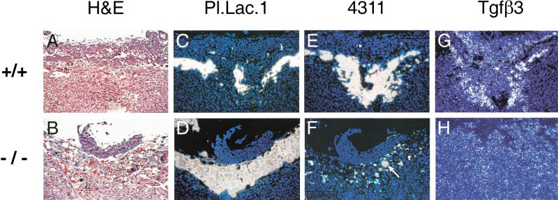
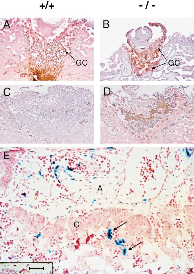
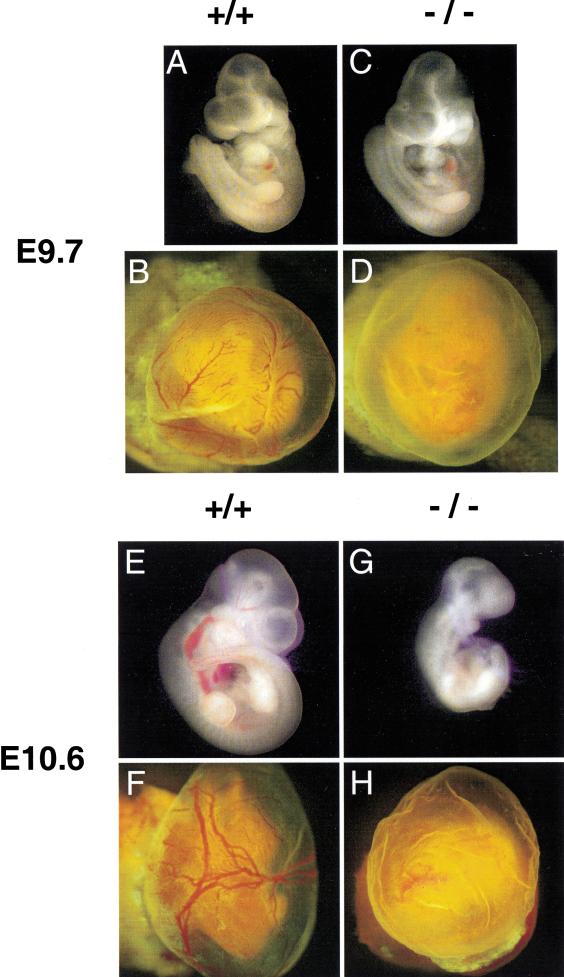
Placental development is profoundly influenced by oxygen (O(2)) tension. Human cytotrophoblasts proliferate in vitro under low O(2) conditions but differentiate at higher O(2) levels, mimicking the developmental transition they undergo as they invade the placental bed to establish the maternal-fetal circulation in vivo. Hypoxia-inducible factor-1 (HIF-1), consisting of HIF-1alpha and ARNT subunits, activates many genes involved in the cellular and organismal response to O(2) deprivation. Analysis of Arnt(-/-) placentas reveals an aberrant cellular architecture due to altered cell fate determination of Arnt(-/-) trophoblasts. Specifically, Arnt(-/-) placentas show greatly reduced labyrinthine and spongiotrophoblast layers, and increased numbers of giant cells. We further show that hypoxia promotes the in vitro differentiation of trophoblast stem cells into spongiotrophoblasts as opposed to giant cells. Our results clearly establish that O(2) levels regulate cell fate determination in vivo and that HIF is essential for mammalian placentation. The unique placental phenotype of Arnt(-/-) animals also provides an important tool for studying the disease of preeclampsia. Interestingly, aggregation of Arnt(-/-) embryonic stem (ES) cells with tetraploid wild-type embryos rescues their placental defects; however, these embryos still die from yolk sac vascular and cardiac defects.


Figures

Figure 1
Figure 1
Vascular defects in E9.5 Arnt−/− placentas. H&E-stained Arnt+/+ (A,B) and Arnt−/− (C,D) sections showing the presence of a chorionic plate (short arrows in A and C) in both animals, but lack of fetal vessels (long arrows in B) in the labyrinthine layer of the Arnt−/− placenta (D). Original magnification in A,C: 80×; magnification of inset in B,D: 400×. (GC) Giant cells; (MD) maternal deciduum; (LT) labyrinthine trophoblast.
Figure 2
Figure 2
Presence of cells expressing placental lactogen 1 (Pl.Lac.1), 4311, and TGFβ3 in E8.5 Arnt+/+ and Arnt−/− placentas. (A,B) hematoxylin- and eosin-stained sections from maternal tissues (Arnt+/−) and placentas with the indicated genotypes. (CH) Adjacent sections analyzed via 35S in situ hybridization. Magnification, 100×.
Figure 3
Figure 3
Presence of cells expressing placental lactogen 1 (Pl.Lac.1), 4311, and TGFβ3 in E9.5 Arnt+/+ and Arnt−/− placentas. (AH) Sections treated as described for Figure 2. Note the expanded population of giant cells apparent in the sections presented in B and D, and the decreased TGFβ3 expression in H. Magnification, 100×.
Figure 4
Figure 4
Arnt+/+ and Arnt−/− placental trophoblasts show similar numbers of Ki-67+ proliferating cells at E8.5 (A,B) and E9.5 (C,D) of embryonic development. Paraffin-embedded sections were immunostained with anti–Ki-67 antibodies and counterstained with hematoxylin. The anti–Ki-67 antibody is conjugated with horseradish peroxidase, and Ki-67+ cells appear reddish brown in AD. The majority of placental cell proliferation has stopped by E9.5, as shown in C and D. (E) E9.5 placenta from a chimeric embryo generated with LacZ- tagged Arnt−/− ES cells. Arrows indicate LacZ+ Arnt−/− endothelial cells within the labyrinthine layer of the chimeric placenta. Magnification in A,B: 100×; C,D: 40×; E: 400×. (GC) Giant cells; (A) allantois; (C) chorion.
Figure 5
Figure 5
(A) Proliferation of TS cells during culture for 4 d. Arnt−/− TS cells show reduced proliferation compared with Arnt+/+ TS cells, under both normoxic and hypoxic culture conditions. (B) Northern blot analysis of total RNA derived from TS cells cultured for 4 d in undifferentiating (U) or differentiating (D), as well as normoxic (20% O2) or hypoxic (3% O2) conditions. 4311, a marker for spongiotrophoblasts, is detected in differentiated wild-type cells and is elevated four- to fivefold under hypoxia. In contrast, little if any 4311 can be detected in Arnt−/− cells. ERRβ2, a marker specific for undifferentiated TS cells, is down-regulated in all TS lines on differentiation. (C) RT–PCR analysis for TGFβ3. After 4 d, differentiated wild-type TS cells show elevated TGFβ3 expression at 3% O2. In contrast, Arnt−/− TS cells did not show any change in TGFβ3 expression when cultured under hypoxia.
Figure 6
Figure 6
Rescue of Arnt−/− placental defects by aggregation of Arnt−/−ES cells with tetraploid wild-type morulas that provide functional extraembryonic tissues. (A–D) E9.7 tetraploid embryo ⇔ ES cell chimeras generated with either Arnt+/+ ES cells (A,B) or Arnt−/− ES cells (C,D) and green fluorescent protein–tagged wild-type tetraploid embryos. Yolk sacs are shown in B and D, whereas embryos dissected free of their yolk sacs are shown in A and C. (E–H) E10.6 chimeras generated with Arnt+/+ and Arnt−/− ES cells. Note the abnormal yolk sac vasculature exhibited by Arnt−/− chimeras at both E9.7 (D) and E10.6 (H).
Figure 7
Figure 7
Yolk sac and cardiac defects detected in tetraploid embryo ⇔ Arnt−/− ES cell chimeras. (A,B) Normal labyrinthine layers containing fetal blood vessels (arrows) observed in both Arnt+/+ and Arnt−/− E9.7 chimeric placentas. However, the anomalous yolk sac blood vessel phenotype detected in nonchimeric Arnt−/− animals is preserved in tetraploid embryo ⇔ ES cell chimeras (although all endodermal derivatives are wild type for the Arnt locus), based on H&E-stained (C,D) and lectin-stained (E,F) sections. Arrows in E and F indicate endothelial cells within the yolk sac vessels that bind lectin and stain black in this assay. (G,H) Sections obtained from Arnt+/+ and Arnt−/− chimeric embryos stained with lectin and counterstained with hematoxylin to show abnormal hearts in the Arnt−/− chimeras. Note the diminished endocardial cushions (EC) and hypoplastic myocardium (arrow) of the Arnt−/− heart. Furthermore, vessels such as the dorsal aortae (arrowheads) appear distended, possibly indicating congestive heart failure. (A) Atrium; (EC) endocardial cushion; (V) ventricle. Magnification in A,B,E,F: 400×; C,D: 200×; G,H: 100×.

Similar articles

Cited by

References

    1. Adelman DM, Maltepe E, Simon MC. Multilineage embryonic hematopoiesis requires hypoxic ARNT activity. Genes & Dev. 1999;13:2478–2483. - PMC - PubMed
    1. Akazawa S, Unterman T, Metzger BE. Glucose metabolism in separated embryos and investing membranes during organogenesis in the rat. Metabolism. 1994;43:830–835. - PubMed
    1. An WG, Kanekal M, Simon MC, Maltepe E, Blagosklonny MV, Neckers LM. Stabilization of wild-type p53 by hypoxia-inducible factor 1α. Nature. 1998;392:405–408. - PubMed
    1. Aplin JD. Hypoxia and human placental development. J Clin Invest. 2000;105:559–560. - PMC - PubMed
    1. Bunn HF, Poyton RO. Oxygen sensing and molecular adaptation to hypoxia. Physiol Rev. 1996;76:839–885. - PubMed

Publication types

MeSH terms

Substances

LinkOut - more resources