. 2000 Dec 15;20(24):8996-9003.
doi: 10.1523/JNEUROSCI.20-24-08996.2000.

Water permeability of cochlear outer hair cells: characterization and relationship to electromotility

Affiliations

Water permeability of cochlear outer hair cells: characterization and relationship to electromotility

I A Belyantseva et al. J Neurosci. .

Abstract

The distinguishing feature of the mammalian outer hair cells (OHCs) is to elongate and shorten at acoustic frequencies, when their intracellular potential is changed. This "electromotility" or "electromechanics" depends critically on positive intracellular pressure (turgor), maintained by the inflow of water through yet uncharacterized water pathways. We measured the water volume flow, J(v), across the plasma membrane of isolated guinea pig and rat OHCs after osmotic challenges and estimated the osmotic water permeability coefficient, P(f), to be approximately 10(-2) cm/sec. This value is within the range reported for osmotic flow mediated by the water channel proteins, aquaporins. J(v) was inhibited by HgCl(2), which is known to block aquaporin-mediated water transport. P(f) values that were estimated for OHCs from neonatal rats were of the order of approximately 2 x 10(-3) cm/sec, equivalent to that of membranes lacking water channel proteins. In an immunofluorescence assay we showed that an anti-peptide antibody specific for aquaporins labels the lateral plasma membrane of the OHC in the region in which electromotility is generated. Using patch-clamp recording, we found that water influx into the OHC is regulated by intracellular voltage. We also found that the most pronounced increases of the electromotility-associated charge movement and of the expression of OHC water channels occur between postnatal days 8 and 12, preceding the onset of hearing function in the rat. Our data indicate that electromotility and water transport in OHCs may influence each other structurally and functionally.


Figures
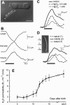
Fig. 1.
Fig. 1.
Characterization of water permeability in OHCs.A, Video image of an OHC from a 12-d-old rat showing the position of the puff pipette relative to the stimulated cell. Scale bar, 10 μm. B, Changes of cell length, diameter, and volume after the pressure application of a hypo-osmotic solution (320 mOsm/kg) to the cell in A. The solid horizontal bar indicates the timing of the solution application. Thedashed line superimposed on the volume trace is a linear fit through the rising phase of the volume response. Line slope measures the speed of the volume increase. C, Volume changes of a different OHC from an adult rat evoked by a hypo-osmotic challenge before (solid line) and during two consecutive bath applications of the water transport inhibitor HgCl2(0.5 mm, dashed line; 1 mm,dotted line). D, Volume changes of an 85-μm-long guinea pig OHC (shown as inset) caused by the application of the hypo-osmotic solution to the apical (dashed line), central (solid line), and basal (dotted line) portions of the cell.Arrows indicate the positions of the puff pipette. Scale bar, 10 μm. E, Developmental changes of the water permeability coefficient (Pf) of OHCs isolated from the apical turn of the rat cochlea. Eachpoint represents the mean ± SE for several cells (4 ≤ n ≤ 8) tested at each particular age.
Fig. 2.
Fig. 2.
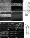
Immunolocalization of the antigen recognized by the pan-aquaporin antibody. A, Bright-field confocal image of the whole mount of the organ of Corti. B, Same confocal plane observed in epifluorescent illumination to show the immunolabeling reaction with the affinity-purified pan-aquaporin antibody. The sample was permeabilized with Triton X-100.C, Conventional fluorescent image of the nonpermeabilized sample immunolabeled with the pan-aquaporin antibody.D, Suppression of immunolabeling after preadsorption of the antibody with the immunizing peptide. E, Western blot analysis of the proteins recognized by the pan-aquaporin antibody in different tissues. Shown from left toright are the organ of Corti from rats of different age (PD0, PD6, adult), kidney, and lens of adult rat. The major ∼58 kDa band is revealed in the organ of Corti (arrow). Multiple bands are seen in other tissues. F, Consecutive confocal optical sections (0.7 μm thickness, numbered 1–6) taken at 2.8, 4.2, and 5.6 μm below the cuticular plate (top row) and at 4.9, 3.5, and 2.1 μm from the base of the cell (bottom row). At intermediate positions along the length of the cell the antibody distinctly labeled the lateral wall of the OHCs, producing annular fluorescence patterns similar to those shown in panels3 and 4. G, Schematic view of confocal sections shown in F. Scale bars, 10 μm.
Fig. 3.
Fig. 3.
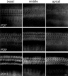
Postnatal development of protein expression revealed by the pan-aquaporin antibody. Organs of Corti of 7-d-old (PD7; toprow), 9-d-old (PD9; middle row), and 12-d-old (PD12; bottom row) rats were separated into three segments (basal, middle, andapical) and processed simultaneously. Scale bar, 10 μm.
Fig. 4.
Fig. 4.
Postnatal growth of the electromotile responses and motility-associated charge movement in OHCs of the apical turn of the rat's organ of Corti. A, Voltage dependence of the specific nonlinear capacitance (in μF/cm2) for two sample cells from the apex of the cochlea at PD5 (open squares) and PD12 (closed circles). Data are fit with the derivative of a Boltzmann function. B, OHC length changes versus transmembrane voltage in the percentage of the cell length at the holding potential (−60 mV) for the same cells that are shown in A. Data are fit with a Boltzmann function.C, Density of the motility-associated charge movement (e/μm2) versus days after birth (mean ± SE). The number of cells is shown above each point.
Fig. 5.
Fig. 5.
Voltage dependence of water permeability.A, Video image of a guinea pig OHC that was patch-clamped at the basal pole with the puff pipette, containing the hypo-osmotic solution, situated 25–30 μm from the cell. Scale bar, 10 μm. B, Representative data set showing, fromtop to bottom, voltage steps delivered through the patch pipette (V; driving voltages indicated near the traces), whole-cell current (I), length changes (L), and volume changes (Volume). At 5 sec after delivery of a voltage step the cell was subjected to the standard hypo-osmotic challenge (320 mOsm/kg; solid horizontal bars). Different line types designate length and volume changes at different step potentials: −90 mV, solid lines; −30 mV, dashed lines; +60 mV, dotted lines.C, Osmotic water flow (Jv) versus membrane potential. After correction of the potentials for the voltage drop across access resistance, Jv measurements in five cells were normalized to the area of the lateral plasma membrane (see Materials and Methods), grouped in 30 mV intervals starting from −80 mV, and averaged. Each point represents the mean ± SE (4 ≤ n ≤ 5).

References

    1. Beitz E, Kumagami H, Krippeit-Drews P, Ruppersberg JP, Schultz JE. Expression pattern of aquaporin water channels in the inner ear of the rat. The molecular basis for a water regulation system in the endolymphatic sac. Hear Res. 1999;132:76–84. - PubMed
    1. Belyantseva IA, Frolenkov GI, Streett D, Wade J, Kachar B. Is the voltage-driven motor protein for outer hair cell electromotility a member of the aquaporin protein family? Abstracts of 22nd Meeting of the Association for Research in Otolaryngology Popelka GR. 1999, abstract 731. St. Petersburg Beach, FL: ARO.
    1. Brown D, Katsura T, Kawashima M, Verkman AS, Sabolic I. Cellular distribution of the aquaporins: a family of water channel proteins. Histochem Cell Biol. 1995;104:1–9. - PubMed
    1. Brownell WE. Outer hair cell electromotility and otoacoustic emissions. Ear Hear. 1990;11:82–92. - PMC - PubMed
    1. Butkus A, Alcorn D, Earnest L, Moritz K, Giles M, Wintour EM. Expression of aquaporin-1 (AQP1) in the adult and developing sheep kidney. Biol Cell. 1997;89:313–320. - PubMed

Publication types

MeSH terms

LinkOut - more resources