. 2001 Feb;68(2):313-24.
doi: 10.1086/318207. Epub 2000 Dec 20.

Wild-type huntingtin reduces the cellular toxicity of mutant huntingtin in vivo

Affiliations

Wild-type huntingtin reduces the cellular toxicity of mutant huntingtin in vivo

B R Leavitt et al. Am J Hum Genet. 2001 Feb.

Abstract

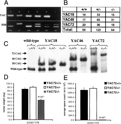
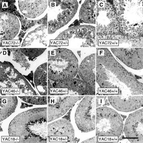
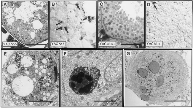
We have developed yeast artificial chromosome (YAC) transgenic mice expressing normal (YAC18) and mutant (YAC46 or YAC72) human huntingtin (htt), in a developmental- and tissue-specific manner, that is identical to endogenous htt. YAC72 mice develop selective degeneration of medium spiny projection neurons in the lateral striatum, similar to what is observed in Huntington disease. Mutant human htt expressed by YAC transgenes can compensate for the absence of endogenous htt and can rescue the embryonic lethality that characterizes mice homozygous for targeted disruption of the endogenous Hdh gene (-/-). YAC72 mice lacking endogenous htt (YAC72 -/-) manifest a novel phenotype characterized by infertility, testicular atrophy, aspermia, and massive apoptotic cell death in the testes. The testicular cell death in YAC72 -/- mice can be markedly reduced by increasing endogenous htt levels. YAC72 mice with equivalent levels of both wild-type and mutant htt (YAC72 +/+) breed normally and have no evidence of increased testicular cell death. Similar findings are seen in YAC46 -/- mice compared with YAC46 +/+ mice, in which wild-type htt can completely counteract the proapoptotic effects of mutant htt. YAC18 -/- mice display no evidence of increased cellular apoptosis, even in the complete absence of endogenous htt, demonstrating that the massive cellular apoptosis observed in YAC46 -/- mice and YAC72 -/- mice is polyglutamine-mediated toxicity from the mutant transgene. These data provide the first direct in vivo evidence of a role for wild-type htt in decreasing the cellular toxicity of mutant htt.


Figures

Figure  1
Figure 1
Rescue of the Hdh-nullizygous lethal phenotype by YAC transgenes expressing mutant htt. Resultant genotypes for the F2 offspring of a cross between a YAC72 transgene–positive, Hdh-heterozygous mouse (+, +/− genotype) and a YAC72 transgene–negative, Hdh-heterozygous mouse (−, +/− genotype) are shown (A). The upper PCR bands represent the presence or absence of the YAC transgene, and the lower bands represent the state of the endogenous Hdh gene. The mouse represented in the second lane has the YAC72 transgene but lacks the endogenous Hdh gene (+, −/− genotype). This mouse demonstrates that mutant human htt expression from our YAC transgene rescued the Hdh-nullizygous state. Mice with targeted disruption of the Hdh gene were rescued from the embryonic lethal phenotype equally by all three of the YAC transgenes described in the present study. The F2 offspring of our experimental breedings had the expected 1:2:1 ratio of genotypes for all of the YAC transgenes examined (B). Western blot analysis of htt-protein expression (C) confirmed the absence of endogenous htt protein in Hdh-nullizygous mice (−/−), compared with wild-type mice (+/+), and demonstrated similar levels of human transgenic htt expression in YAC18-, YAC46-, and YAC72-rescued Hdh-nullizygous mice (+, −/−). Average testicular weight (D) and epididymal sperm counts (E) for YAC72 +/+ mice, YAC72 +/− mice, and YAC72 −/− mice at age 4 mo are shown. YAC72 −/− mice had significant testicular atrophy (P<10-5) and decreased sperm counts (P<10-5), compared with YAC72 +/+ mice and YAC72 +/− mice.
Figure  2
Figure 2
Testicular morphology of YAC transgene–rescued Hdh-nullizygous mice. Semithin sections of testes stained with toluidine blue that are from 8-mo-old mice reveal the gross testicular morphology of mice with the YAC72 (AC), YAC46 (DF), and YAC18 (GI) HD transgenes and either complete absence of endogenous htt (−/−), 50% of endogenous htt levels (+/−), or 100% of endogenous htt levels (+/+). Massive degeneration of spermatogenic cells occurs in the seminiferous tubules of mice expressing mutant htt with either 46 or 72 polyglutamine repeats (A and D). The cell death is most pronounced in YAC72 −/− (A), intermediate in YAC46 −/− (D), and not present in YAC18 −/− (G) Hdh-nullizygous mice. The human HD transgene in each of these lines of mice is identical except for the length of the CAG repeat, and these results suggest that this novel cell-death phenotype is CAG-repeat-length dependent. Increasing levels of endogenous htt markedly reduced the amount of spermatogenic-cell degeneration (B, C, E, and F) observed in YAC46 and YAC72 mice. (Scale bar = 100 μm)
Figure  3
Figure 3
Morphological, biochemical, and ultrastructural evidence for apoptotic cell death in the testes of YAC72 mice lacking endogenous htt. Toluidine blue staining revealed massive death of spermatogenic cells in the testes of YAC72 −/− mice. Decreased numbers of spermatogenic cells and a disordered epithelium filled with vacuoles are present in these testes, compared with the very large numbers of spermatocytes in the well-ordered stratified epithelium of testes of YAC72 +/+ mice. Increased apoptosis was evident in the testes of YAC72 −/− mice, on the basis of increased TUNEL labeling of spermatogenic cells (B, arrows), compared with YAC72 mice that had normal levels of endogenous htt (D). EM analysis of degenerating testicular cells from YAC72 −/− mice also provided evidence of apoptosis. Ultrastructural analysis of testes from Hdh- nullizygous YAC72 mice reveals massive cell death of spermatids, phagocytosis of degenerating cells, and formation of multinucleated giant cells. The epithelium of YAC72 mice lacking endogenous htt was characterized by degenerating spermatids filled with cytoplasmic vacuoles (E), phagosomes containing shrunken electron-dense spermatids engulfed within Sertoli cells (F), and spermatogenic giant cells (G). (Scale bars in AD = 100 μm; scale bar in E = 10 μm; scale bar in F = 5 μm; scale bar in G = 10 μm)
Figure  4
Figure 4
Protein aggregates in YAC72 −/− mice. Ultrastructural analysis of the testes of YAC72 mice lacking endogenous htt revealed the occasional presence of abnormal aggregates of intracellular protein (arrowheads) within elongate spermatids (A), Sertoli cells (B), and sperm tails (C). The composition of these protein aggregates is not entirely clear, but they resemble the ultrastructural appearance of htt aggregates found in human HD brain tissue. Ectopic microtubule bundles (D) and manchettes (E) also were identified (asterisks [*]). The bundle in panel E is in a spermatogonium. N = nucleus. (Scale bars in A and B = 5 μm; scale bar in C and D = 1 μm; scale bar in E = 5 μm)
Figure  5
Figure 5
Immunocytochemical analysis of protein aggregates and actin distribution in sections from the testes of YAC72 −/− mice. Abnormal protein aggregates within degenerating spermatogenic cells in the testes of YAC72 −/− mice contain htt (phase [A] and immunofluorescence [B]). In normal epithelium (phase [E] and fluorescence [F]), actin filaments in Sertoli cells are concentrated in unique adhesion plaques (ectoplasmic specializations) that occur at apical sites of attachment to spermatids (Apical) and at basal sites of attachment to neighboring Sertoli cells (Basal). In YAC72 −/− mice (GJ), filament bundles (asterisks [*]) in apical regions occur in areas not associated with spermatid heads, although filament bundles at basal sites occur in their normal position. (Scale bars in AD = 10 μm; scale bars in EJ = 50 μm)

References

Electronic-Database Information

    1. Genome Database, The, http://www.gdb.org (for HD [accession number 119307])
    1. Online Mendelian Inheritance in Man (OMIM), http://www.ncbi.nlm.nih.gov/Omim/ (for HD [MIM 143100])

References

    1. Andrew SE, Goldberg YP, Kremer B, Telenius H, Theilmann J, Adam S, Starr E, Squitieri F, Lin B, Kalchman MA, Graham RK, Hayden MR (1993) The relationship between trinucleotide (CAG) repeat length and clinical features of Huntington's disease. Nat Genet 4:398–403 - PubMed
    1. Bao J, Sharp AH, Wagster MV, Becher M, Schilling G, Ross CA, Dawson VL, Dawson TM (1996) Expansion of polyglutamine repeat in huntingtin leads to abnormal protein interactions involving calmodulin. Proc Natl Acad Sci USA 93:5037–5042 - PMC - PubMed
    1. Becher MW, Kotzuk JA, Sharp AH, Davies SW, Bates GP, Price DL, Ross CR (1998) Intranuclear neuronal inclusions in Huntington`s disease and dentatorubral and pallidoluysian atrophy: correlation between density of inclusions and IT15 triplet repeat length. Neurobiol Dis 4:387–397 - PubMed
    1. Burke JR, Enghild JJ, Martin ME, Jou Y-S, Myers RM, Roses AD, Vance JM, Strittmatter WJ (1996) Huntingtin and DRPLA proteins selectively interact with the enzyme GADPH. Nat Med 2:347–349 - PubMed
    1. Cooper JK, Schilling G, Peters MF, Herring WJ, Sharp AH, Kaminsky Z, Masone J, Khan FA, Delanoy M, Borschelt DR, Dawson VL, Dawson TM, Ross CA (1998) Truncated N-terminal fragments of huntingtin with expanded glutamine repeats form nuclear and cytoplasmic aggregates in cell culture. Hum Mol Genet 7:783–790 - PubMed

Publication types

MeSH terms

Associated data

LinkOut - more resources