

Review
doi: 10.1136/bjo.85.1.102.
Idiopathic full thickness macular hole: natural history and pathogenesis
Affiliations
- PMID: 11133724
- PMCID: PMC1723702
- DOI: 10.1136/bjo.85.1.102
Item in Clipboard
Review
Idiopathic full thickness macular hole: natural history and pathogenesis
Br J Ophthalmol.
2001 Jan.
No abstract available
Figures
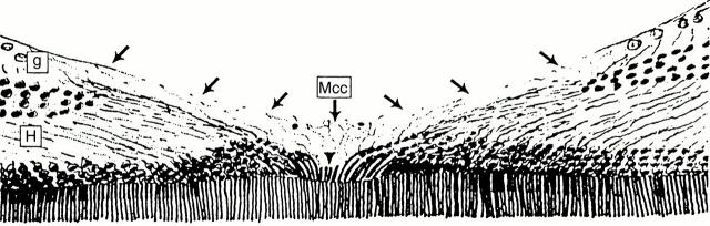
Schematic representation of the normal human fovea centralis from Gass62 based on the histological findings of Yamada63 and Hogan et al.64 The Muller cell cone (Mcc) is shown with its base forming the internal limiting membrane (arrows) and its truncated apex forming the outer limiting membrane at the umbo (arrowhead). The Henle nerve fibre layer (H) and the foveal extremety of the ganglion cell layer (g) are also demonstrated. (From Gass JDM, "The Muller cell cone. An overlooked part of the normal fovea". Arch Ophthalmol 1999;117:821-3. Copyrighted 1999, American Medical Association.)
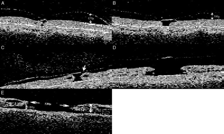
Evolution of idiopathic FTMHs on OCT. A 69 year old female with a long standing FTMH in the first eye and a 2 month history of central visual loss (acuity 6/12) and metamorphopsia in the fellow eye caused by a stage 1a lesion. OCT examination (A) revealed a central foveal detachment and a high reflectance interface in the preretinal plane which is thought to represent the posterior vitreous cortex, partially detached at the macula but remaining tethered to the central fovea. (B) Two months later a stage 1b lesion was noted on funduscopy and OCT revealed a more extensive foveal detachment. (C) At 4 months after the onset of symptoms, she had developed a stage 2 lesion with the acuity dropping to 6/24. The OCT confirms the presence of a pericentric full thickness break (arrow) and the early formation of an operculum. Cystic spaces are present at the edges of the hole. The patient elected to undergo surgical treatment at this stage. (D) An OCT of a stage 3 lesion in a 70 year old woman, showing a FTMH (380 µm) with vitreomacular separation and an operculum suspended on the posterior vitreous face. A prominent subretinal fluid cuff is visible (asterisk). (E) A stage 4 lesion in a 65 year old man. Note the extensive subretinal fluid cuff and the prominent cystoid spaces at the level of the inner plexiform layer at the edges of the hole.
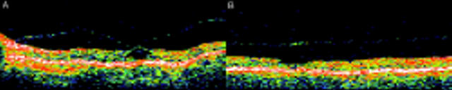
Spontaneous arrest of a stage 1 lesion. (A) OCT of the fellow eye of a 64 year old woman with a stage 1a lesion on funduscopy and acuity of 6/9. The lesion was observed and 2 months later she reported an improvement in her metamorphopsia. Funduscopy revealed a reattached fovea with an inner lamellar defect. A free operculum was present in the preretinal plane with vitreomacular separation but otherwise attached vitreous. OCT (B) confirmed the clinical findings and no residual vitreofoveal traction. The visual acuity remained at 6/9 throughout follow up.

Spontaneous arrest and closure of a stage 2 lesion. (A) OCT of the first eye of a 56 year old female who presented with a stage 2 "can opener" lesion and a visual acuity of 6/36. Three months later she reported an improvement in the visual acuity (6/12) and funduscopy revealed a normal foveal reflex with vitreomacular separation and a free floating operculum. OCT (B) showed a relatively normal foveal contour, vitreomacular separation with an operculum, and no residual vitreofoveal traction.
References
Publication types
MeSH terms
LinkOut - more resources
Full Text Sources