. 2000 Dec 25;151(7):1469-82.
doi: 10.1083/jcb.151.7.1469.

Metaphase to anaphase (mat) transition-defective mutants in Caenorhabditis elegans

Affiliations

Metaphase to anaphase (mat) transition-defective mutants in Caenorhabditis elegans

A Golden et al. J Cell Biol. .

Abstract

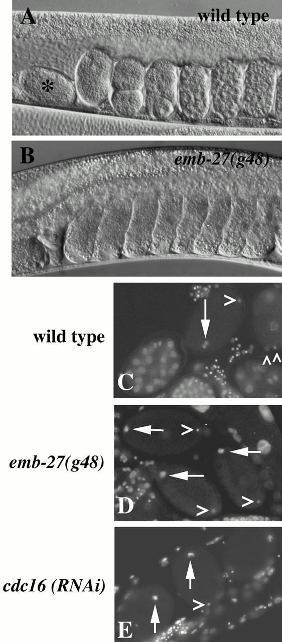
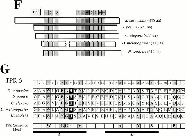
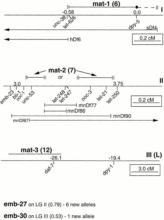
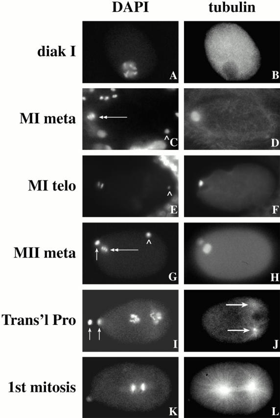
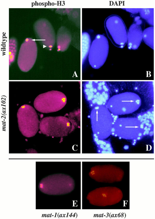
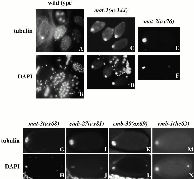
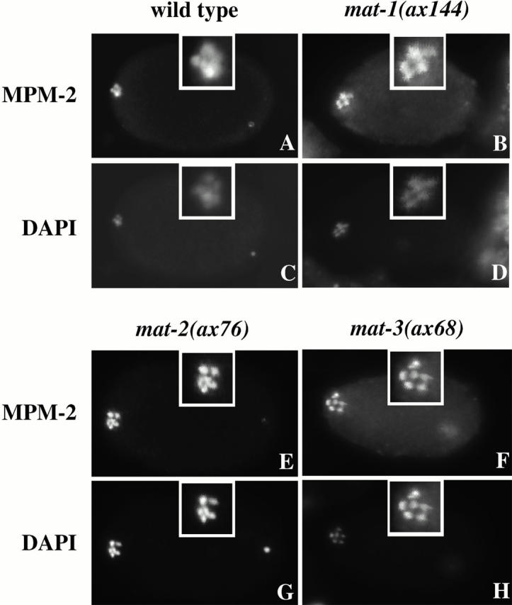
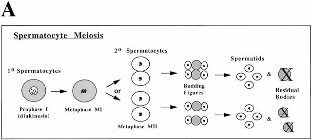
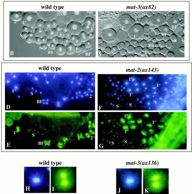
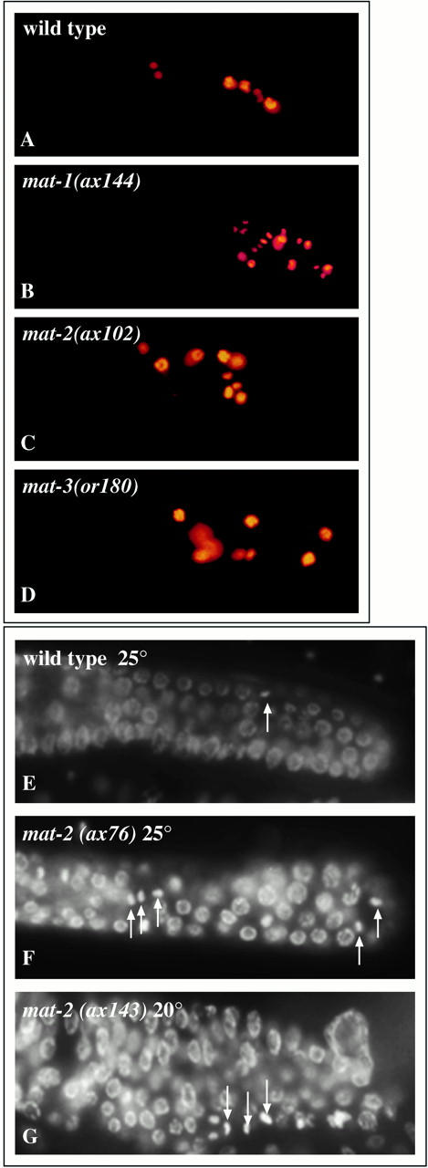
The metaphase to anaphase transition is a critical stage of the eukaryotic cell cycle, and, thus, it is highly regulated. Errors during this transition can lead to chromosome segregation defects and death of the organism. In genetic screens for temperature-sensitive maternal effect embryonic lethal (Mel) mutants, we have identified 32 mutants in the nematode Caenorhabditis elegans in which fertilized embryos arrest as one-cell embryos. In these mutant embryos, the oocyte chromosomes arrest in metaphase of meiosis I without transitioning to anaphase or producing polar bodies. An additional block in M phase exit is evidenced by the failure to form pronuclei and the persistence of phosphohistone H3 and MPM-2 antibody staining. Spermatocyte meiosis is also perturbed; primary spermatocytes arrest in metaphase of meiosis I and fail to produce secondary spermatocytes. Analogous mitotic defects cause M phase delays in mitotic germline proliferation. We have named this class of mutants "mat" for metaphase to anaphase transition defective. These mutants, representing six different complementation groups, all map near genes that encode subunits of the anaphase promoting complex or cyclosome, and, here, we show that one of the genes, emb-27, encodes the C. elegans CDC16 ortholog.


Figures

Figure 1
Figure 1
Molecular genetic analysis of emb-27. Shown are DIC micrographs of embryos within the uteri of wild-type (A) and emb-27(g48) (B) hermaphrodites. Oocytes are fertilized in the spermatheca and pass into the uterus one by one. In the wild-type hermaphrodite (A), the horizontally positioned embryo within the spermatheca (*) is a meiotic one-cell embryo. Within the uterus are (left to right) a mitotic one-cell, two-cell, and additional multicellular embryos. (B) The emb-27(g48) adult hermaphrodite was shifted to 25°C as an L1 larva. emb-27 mothers contain only one-cell embryos. Note, ventral is down. (C–E) DAPI-stained embryos isolated from the uteri of young adult hermaphrodites. White arrows denote clusters of oocyte chromosomes, and > mark the single masses of hypercondensed sperm chromatin. Wild-type spreads (C) include embryos of many different ages. In the central, meiotic one-cell embryo, the sperm chromosomes are hypercondensed, and the oocyte chromosomes are in anaphase of meiosis I. No polar bodies are present. In contrast, the mitotic one-cell embryo on the right has extruded two polar bodies (marked by Λ), and its oocyte and sperm pronuclei are in mitotic prophase. emb-27(g48) spreads (D) contain only one-cell embryos. Each of the three embryos shown lacks polar bodies and contains a mass of hypercondensed sperm chromatin and a single array of oocyte chromosomes. cdc16(RNAi) embryos (E) phenocopy those from emb-27(g48) mothers. Embryos in this figure and in all subsequent figures are ∼50 μm in length. (F) The distribution of TPR-repeat motifs within C. elegans F10B5.6 is similar to that of cdc16 genes from other species and different from that of other TPR-containing proteins. cdc16 sequences were compared and overlaid using the Clustal W multiple sequence alignment program. TPR5 and TPR6 (darkened) exhibit the highest degree of cross-species amino acid sequence identity. An extended region of the Drosophila melanogaster sequence has been deleted to emphasize the relative alignment of the TPR motifs. (G) Sequence alignment of TPR6. Each TPR consists of 34 amino acids; the conserved hydrophobic residues are shaded in light gray. These residues lie on the hydrophobic faces of the two amphipathic α helices (residues 1–12 and 17–28; A and B, respectively) (Blatch and Lassle 1999). The asterisk at position 9 denotes the conserved histidine (shaded in black) that is altered to a tyrosine in emb-27(g48). The sequence data for the wild-type emb-27 gene is available from GenBank/EMBL/DDBJ under accession no. AF314467.
Figure 1
Figure 1
Molecular genetic analysis of emb-27. Shown are DIC micrographs of embryos within the uteri of wild-type (A) and emb-27(g48) (B) hermaphrodites. Oocytes are fertilized in the spermatheca and pass into the uterus one by one. In the wild-type hermaphrodite (A), the horizontally positioned embryo within the spermatheca (*) is a meiotic one-cell embryo. Within the uterus are (left to right) a mitotic one-cell, two-cell, and additional multicellular embryos. (B) The emb-27(g48) adult hermaphrodite was shifted to 25°C as an L1 larva. emb-27 mothers contain only one-cell embryos. Note, ventral is down. (C–E) DAPI-stained embryos isolated from the uteri of young adult hermaphrodites. White arrows denote clusters of oocyte chromosomes, and > mark the single masses of hypercondensed sperm chromatin. Wild-type spreads (C) include embryos of many different ages. In the central, meiotic one-cell embryo, the sperm chromosomes are hypercondensed, and the oocyte chromosomes are in anaphase of meiosis I. No polar bodies are present. In contrast, the mitotic one-cell embryo on the right has extruded two polar bodies (marked by Λ), and its oocyte and sperm pronuclei are in mitotic prophase. emb-27(g48) spreads (D) contain only one-cell embryos. Each of the three embryos shown lacks polar bodies and contains a mass of hypercondensed sperm chromatin and a single array of oocyte chromosomes. cdc16(RNAi) embryos (E) phenocopy those from emb-27(g48) mothers. Embryos in this figure and in all subsequent figures are ∼50 μm in length. (F) The distribution of TPR-repeat motifs within C. elegans F10B5.6 is similar to that of cdc16 genes from other species and different from that of other TPR-containing proteins. cdc16 sequences were compared and overlaid using the Clustal W multiple sequence alignment program. TPR5 and TPR6 (darkened) exhibit the highest degree of cross-species amino acid sequence identity. An extended region of the Drosophila melanogaster sequence has been deleted to emphasize the relative alignment of the TPR motifs. (G) Sequence alignment of TPR6. Each TPR consists of 34 amino acids; the conserved hydrophobic residues are shaded in light gray. These residues lie on the hydrophobic faces of the two amphipathic α helices (residues 1–12 and 17–28; A and B, respectively) (Blatch and Lassle 1999). The asterisk at position 9 denotes the conserved histidine (shaded in black) that is altered to a tyrosine in emb-27(g48). The sequence data for the wild-type emb-27 gene is available from GenBank/EMBL/DDBJ under accession no. AF314467.
Figure 2
Figure 2
Genetic map of the mat mutants and emb-27 and emb-30. See Materials and Methods for genetic details.
Figure 3
Figure 3
Tubulin and DAPI localization during meiotic progression of wild-type oocytes before and after fertilization. Shown in A, C, E, G, I, and K are DAPI images, and in B, D, F, H, J, and L are the corresponding tubulin images. An oocyte in diakinesis of meiotic prophase I (diak I) is shown in A and B. The subsequent stages after fertilization are depicted: meiosis I metaphase (C and D, MI meta), meiosis I telophase (E and F, MI telo), meiosis II metaphase (G and H, MII meta), transitional prophase (I and J, Trans'l Pro) of the first mitotic cell cycle, and then first mitosis (K and L). During metaphase of meiosis I, the six bivalents are often distinguishable as a “pentagonal array” or, in side view, as a cluster of three axially aligned bivalents (C). During rotation, chromosomes enter anaphase and often up to 12 distinguishable homologues can be visualized at this time. By the time rotation is complete (E and F), the homologues have moved apart towards the opposite spindle poles. Univalents are no longer distinguishable. The oocyte meiotic chromosomes are indicated by double arrows (C and G). Polar bodies, the discarded meiotic products, are indicated by vertical arrows (G and I). In C, E, and G, the condensed sperm chromatin (marked with Λ) marks the future embryonic posterior. The duplicated sperm asters are indicated by arrows (J). C. elegans embryos are ∼50 μm in length.
Figure 5
Figure 5
Phosphohistone H3 and DAPI localization in one-cell arrested mat embryos. Embryos were dissected from mothers that had been upshifted to 25°C for 6–7 h (A and B). The three wild-type, one-cell stage embryos are in meiosis I metaphase (left), meiosis II metaphase (middle), and S phase (right), based on their DNA staining patterns (B). In the wild-type panels, phosphohistone H3 stains the pentagonal array of the oocyte metaphase meiosis I chromosomes (arrow), but it does not stain the sperm chromatin mass on the other side of the embryo. In the middle embryo, the sperm chromatin mass is out of focus. Phosphohistone H3 brightly stains both the oocyte meiosis II chromosomes (arrowhead) and the chromatin within the adjacent polar body. In the S phase embryo, phosphohistone H3 stains the two polar bodies and the oocyte pronucleus, but not the male pronucleus, which, in this particular embryo, lies next to the oocyte pronucleus. All three mat-2(ax102) embryos are in metaphase of meiosis I (D), and their congressed chromosomes (D, arrows) stain brightly (C). Congressed chromosomes also stain in mat-1(ax144) (E) and mat-3(ax68) (F). (F) The bottom mat-3 embryo is older and displays the terminal phenotype; the chromosomes continue to stain with phospho-H3 antibody, but have moved centrally and lost their condensed metaphase chromosome morphology. The out of focus DAPI-stained body in the top embryo in D is outside of the embryo.
Figure 4
Figure 4
Tubulin and DAPI localization in one-cell arrested mat embryos. Wild-type and mat mutant adults were shifted to 25.5°C for 6–7 h before embryos were dissected from their mothers. Shown in A and B are a spread of embryos from a wild-type mother. Wild-type animals display a range of developmental stages from meiotic and mitotic one-cells to multicellular. An embryo in metaphase of meiosis I is visible on the left in A; an arrow marks its chromosomes in B. Shown in C and D are a spread of embryos from a mat-1(ax144) mother in which all the embryos are arrested in metaphase of meiosis I. E–N show single embryos stained with an anti-tubulin antibody (E, G, I, K, and M). The corresponding DAPI image is shown below each tubulin image. Anterior is to the left. Of these mutants, only emb-1(hc62) was not identified in our genetic screens. In most images, the hypercondensed sperm chromatin mass is visible at the right end of each embryo.
Figure 6
Figure 6
MPM-2 and DAPI localization in one-cell arrested mat embryos. Embryos were dissected from mothers that had been shifted to 25°C for 6–7 h. These embryos were stained with the MPM-2 monoclonal antibody (A, B, E, and F) and DAPI (C, D, G, and H) to identify congressed M phase–like chromosomes (A, B, E, and F). Wild-type embryos in metaphase of meiosis I (A and C) are shown compared with arrested mat-1 (B and D), mat-2 (E and G), and mat-3 (F and H) embryos. Note, anterior is to the left. In most images, the hypercondensed sperm chromatin mass is visible at the right end of each embryo. A magnified view of the oocyte chromosomes are shown in each panel.
Figure 7
Figure 7
Tubulin and DAPI localization in wild-type and mutant spermatocytes. Shown in A is a depiction of wild-type spermatogenesis. After nuclear envelope breakdown, the chromosomes of primary (1°) spermatocytes congress to form a meiosis I metaphase plate. The first meiotic division yields two secondary (2°) spermatocytes, which can either be separated or remain attached through a cytoplasmic connection due to incomplete cytokinesis. Secondary spermatocytes subsequently undergo a polarized budding division during which two haploid sperm separate from a central residual body. (B) A wild-type male germline squashed in the presence of Hoechst dye 33342 and observed by DIC and UV epifluorescence. All meiotic stages can be seen. Primaries (1°) and secondaries (2°) can be distinguished by size (B). A budding figure (BF) is visible in the lower right corner. Haploid sperm (S) and residual bodies (RB) are also indicated. (C) A sperm spread from a mat-3(ax82) male lacks 2° spermatocytes, but contains many anucleate sperm. Abnormal budding figures are also present in which all the chromosomes remain in the center of the developing residual body. Staining with anti–α-tubulin antibody (E, G, I, and K) and DAPI (D, F, H, and L) reveals the underlying spindle structures in these sperm spreads (E, G, I, and K). In wild-type sperm spreads, distinctive meiotic spindles are apparent in 1° and 2° spermatocytes and in budding figures (D and E). A magnified view of a wild-type meiotic spindle is shown in (I) with its corresponding DAPI image (H). In the mat mutants, normal meiosis I–like spindles form, but anaphase figures are never observed (F, G, J, and K). The diameter of a primary spermatocyte equals 12 μm.
Figure 7
Figure 7
Tubulin and DAPI localization in wild-type and mutant spermatocytes. Shown in A is a depiction of wild-type spermatogenesis. After nuclear envelope breakdown, the chromosomes of primary (1°) spermatocytes congress to form a meiosis I metaphase plate. The first meiotic division yields two secondary (2°) spermatocytes, which can either be separated or remain attached through a cytoplasmic connection due to incomplete cytokinesis. Secondary spermatocytes subsequently undergo a polarized budding division during which two haploid sperm separate from a central residual body. (B) A wild-type male germline squashed in the presence of Hoechst dye 33342 and observed by DIC and UV epifluorescence. All meiotic stages can be seen. Primaries (1°) and secondaries (2°) can be distinguished by size (B). A budding figure (BF) is visible in the lower right corner. Haploid sperm (S) and residual bodies (RB) are also indicated. (C) A sperm spread from a mat-3(ax82) male lacks 2° spermatocytes, but contains many anucleate sperm. Abnormal budding figures are also present in which all the chromosomes remain in the center of the developing residual body. Staining with anti–α-tubulin antibody (E, G, I, and K) and DAPI (D, F, H, and L) reveals the underlying spindle structures in these sperm spreads (E, G, I, and K). In wild-type sperm spreads, distinctive meiotic spindles are apparent in 1° and 2° spermatocytes and in budding figures (D and E). A magnified view of a wild-type meiotic spindle is shown in (I) with its corresponding DAPI image (H). In the mat mutants, normal meiosis I–like spindles form, but anaphase figures are never observed (F, G, J, and K). The diameter of a primary spermatocyte equals 12 μm.
Figure 8
Figure 8
Phospho-H3 staining of adult upshifted wild-type (A), mat-1(ax144) (B), mat-2(ax102) (C), and mat-3(or180) (D) hermaphrodites reveals an excess of mitotic plates in the mitotic region of germlines from isolated mutant gonads. DAPI staining also reveals fewer mitotic figures in the wild-type (E) than mat-2(ax76) (F) or mat-2(ax143) (G) gonads. Some phenotypes were more obvious at an intermediate temperature of 20°C (G).

Similar articles

Cited by

References

    1. Albertson D.G. Formation of the first cleavage spindle in nematode embryos. Dev. Biol. 1984;101:61–72. - PubMed
    1. Albertson D.G., Thomson J.N. Segregation of holocentric chromosomes at meiosis in the nematode, Caenorhabditis elegans . Chromosome Res. 1993;1:15–26. - PubMed
    1. Altschul S.F., Gish W., Miller W., Myers E.W., Lipman D.J. Basic local alignment search tool. J. Mol. Biol. 1990;215:403–410. - PubMed
    1. Beanan M.J., Strome S. Characterization of a germ-line proliferation mutation in C. elegans . Development. 1992;116:755–766. - PubMed
    1. Blatch G.L., Lassle M. The tetratricopeptide repeata structural motif mediating protein-protein interactions. Bioessays. 1999;21:932–939. - PubMed

Publication types

MeSH terms