. 2000 Dec 25;151(7):1483-500.
doi: 10.1083/jcb.151.7.1483.

Flightin is essential for thick filament assembly and sarcomere stability in Drosophila flight muscles

Affiliations

Flightin is essential for thick filament assembly and sarcomere stability in Drosophila flight muscles

M C Reedy et al. J Cell Biol. .

Abstract

Flightin is a multiply phosphorylated, 20-kD myofibrillar protein found in Drosophila indirect flight muscles (IFM). Previous work suggests that flightin plays an essential, as yet undefined, role in normal sarcomere structure and contractile activity. Here we show that flightin is associated with thick filaments where it is likely to interact with the myosin rod. We have created a null mutation for flightin, fln(0), that results in loss of flight ability but has no effect on fecundity or viability. Electron microscopy comparing pupa and adult fln(0) IFM shows that sarcomeres, and thick and thin filaments in pupal IFM, are 25-30% longer than in wild type. fln(0) fibers are abnormally wavy, but sarcomere and myotendon structure in pupa are otherwise normal. Within the first 5 h of adult life and beginning of contractile activity, IFM fibers become disrupted as thick filaments and sarcomeres are variably shortened, and myofibrils are ruptured at the myotendon junction. Unusual empty pockets and granular material interrupt the filament lattice of adult fln(0) sarcomeres. Site-specific cleavage of myosin heavy chain occurs during this period. That myosin is cleaved in the absence of flightin is consistent with the immunolocalization of flightin on the thick filament and biochemical and genetic evidence suggesting it is associated with the myosin rod. Our results indicate that flightin is required for the establishment of normal thick filament length during late pupal development and thick filament stability in adult after initiation of contractile activity.


Figures

Figure 1
Figure 1
Flightin cofractionates with myosin after high salt extraction of skinned IFM fibers. A 10% SDS-PAGE of proteins from a high ionic strength extraction and fractionation of skinned IFM fibers from wild-type flies (MHC+) and from Y97 transgenic flies (MHCY97) expressing a truncated MHC lacking the motor domain (Cripps et al. 1999). F shows total myofibrillar proteins from a skinned fiber before extraction, and P shows the 1-M KCl insoluble pellet after ultracentrifugation at 100,000 g. The supernatant from the 1-M KCl extraction was then diluted 10-fold and subjected to a second ultracentrifugation at 100,000 g. S shows the supernatant and M shows the precipitated, myosin-enriched fraction, which includes MHC, RLC, phosphorylated RLC, ELC, and flightin (FLN). By immunoblot analysis, flightin is detected in the M fraction, but not in the P or S fractions (not shown). Note that flightin is present in the thick filament-enriched M fraction from IFM of Y97 and, as expected, the ELC is absent. The difference in flightin mobility is due to phosphorylation (Vigoreaux et al. 1993). These results suggest that flightin is associated with the thick filament shaft.
Figure 2
Figure 2
The distribution of flightin in myofibrils of wild type and the actin null, KM88. Thin Lowicryl longitudinal sections of Drosophila IFM labeled with anti–flightin polyclonal antibody and Protein A gold. Both sections show tissue from adults >2 d old. (A) Wild type shows labeling on the A band with gaps each side of the Z band and at the H zone. The histogram shows the distribution of gold particles measured in five sarcomeres from different myofibrils. (B) KM88 actin null, where no thin filaments are present, has labeling on bundles of thick filaments. The resolution of the probe is not sufficient to distinguish between label on or between thick filaments, but the presence of flightin in the actin null suggests flightin is associated with the thick filaments. Scale bars: 0.5 μm.
Figure 5
Figure 5
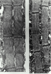
Flightin null assembles long myofilaments and sarcomeres during pupal development. EMs of thin longitudinal sections of IFM from wild-type (left) and fln0 (right) late stage pupa. At this developmental stage, wild-type sarcomeres show an average length of 3.1 μm, while fln0 sarcomeres show an average length of 3.8 μm and are narrower. Note the presence of two transverse electron dense stripes flanking the M line (arrows). These stripes are reminiscent of the transient, particle-dense stripes that have been observed in some late pupa of wild-type IFM (Reedy and Beall 1993). In normal flies, the double band is present for a few hours just before the end of pupation; in fln0 flies, the double band sometimes persists into adulthood.
Figure 3
Figure 3
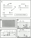
Generation and characterization of a null allele of flightin. (A) Summary of genetic scheme to isolate null mutation in the flightin gene. Male flies fed the mutagen EMS were mated to females heterozygous for Df(3L)fln and the hemizygous progeny were scored for the absence of flightin by immunodot blots. (B) A representative dot blot. The sample encircled, 2987, did not react with an antiflightin mAb. (C) Strain 2987 does not express flightin. Western blot showing the accumulation of α-actinin (top) and flightin (bottom) in IFM of the following strains: (1) Oregon R (wild-type); (2) Df(3L)kto1/+, a deficiency of the 76BD region that does not delete the flightin gene; (3) Df(3L)fln/+; (4) 2987/+; (5) 2987/Df(3L)fln; (6) 2987/2987; (7) +/+ pupa. Note that flightin is not detected in 2987 homozygotes nor 2987/Df(3L)fln transheterozygotes. (D) DNA sequence analysis of the flightin gene from wild-type flies (top) and 2987 (bottom). Note the G to A transition converts a Trp codon at the eighth amino acid position into a stop codon. Herein we will refer to line 2987 as fln0.
Figure 4
Figure 4
Absence of flightin results in large changes in IFM morphology. Scanning electron micrographs of entire half-thoraces from wild-type adult (A), newly eclosed (<30 min) fln0adult (B), and 3-d-old fln0adult (C). In wild-type flies, DLM fibers (*) extend the entire antero-posterior length of the thoracic cavity (A). In contrast, newly eclosed fln0adults occasionally show “wavy” fibers that are longer than normal (arrow). In C, all DLM fibers have detached from the posterior cuticle (arrow). For all figures, anterior is right and dorsal is top. Magnification 150× (A and B) or 200× (C). (d) Time course of fln0fiber degeneration. Flies aged for the indicated times were dissected and their DLM fibers were scored on an arbitrary scale of 0 (normal) to 3 (severely shortened). Each time point represents at least five flies (mean ± SEM). Note that by 8 h, all fibers appear completely degenerated. A similar abnormality has been described for Mhc13(Kronert et al. 1995).
Figure 6
Figure 6
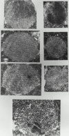
Electron micrographs of cross sections show normal sarcomere assembly of pupal IFM in fln0 and the disruption of adult sarcomeres during the first hours after eclosion. (A–C) Late pupal wild-type (D–F) late pupal fln0, and (G) adult fln0 IFM. Wild-type shows (A) an ordered lattice in Z bands, (B) hollow thick filaments in the A band in a regular hexagonal lattice with thin filaments, and (C) solid thick filament profiles in the M band. fln0 late pupa, although smaller in diameter due to fewer filaments, show normal Z band (D), hollow thick filaments in the A band (E), and solid thick filaments in the M band (F). In adult fln0(G), all sarcomere levels are seen within one section of a single myofibril due to filament misregistration and fragmentation of the sarcomeres. Z bands (Z), A band (A), and M band (M).
Figure 7
Figure 7
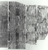
Rapid, progressive sarcomere degeneration in fln0 adult IFM. Electron micrographs of (A) wild-type sarcomeres (B–D) fln0 sarcomeres along the same myofibril in longitudinal 25-nm sections of adult IFM within 12 h after eclosion. In contrast to the relatively well-ordered sarcomeres in pupa (see Fig. 5), adult fln0 sarcomeres are severely disordered. There appears to be a gradual increase in disorder toward one end of a fiber. (B) Even in the best-ordered fln0adult sarcomeres, the M line has disappeared and the Z band tends to fragment. Peripheral bundles of thin filaments (arrowhead) and very long thick filaments (∼10 μm, arrows) are evident. These well-ordered sarcomeres have already shortened to ∼3.1-μm long, about the same length as wild type. (C) Same myofibril as B, but ∼50 μm closer to one end. Note increase in disorder. Z bands are more fragmented and sarcomeres are shorter (∼2.0 μm). In some regions, thick filaments are missing or appear fragmented. Thin filament “cowlicks” project out of the sarcomere (arrowhead). (D) Even more disordered sarcomeres are found further along the same myofibril.
Figure 8
Figure 8
Unusual accumulation of particles in degenerating fln0adult IFM. Longitudinal 60-nm sections of adult IFM. (A) Note abundance of dense particles, similar to those observed in pupa but never seen in wild-type adults. The particles form a “flame-stitch” pattern across the myofibril, usually centered about the former M line (large arrows). (B) Blank gaps in the filament lattice containing larger globular particles (small arrows) are common at this stage. (C) Z bands have broken apart into Z bodies that are no longer in axial register. Spacing between Z bodies is ∼1.4 μm. Regions with numerous thin filaments but devoid of thick filaments also are seen (arrowhead).
Figure 9
Figure 9
Absence of flightin does not impair rigor crossbridge formation. Rigor was induced in glycerinated adult fln 0 IFM by ATP washout. Thin filaments appear decorated throughout by myosin but rigor chevrons (arrowheads) can be seen pointing towards the missing M line only in well-ordered areas. The level of the missing M line is indicated by a transverse arrow. Arrows on thin filaments are ahead of a series of crossbridges on a thin filament and indicate the direction in which the crossbridge arrowheads are pointing. In normal IFM, all rigor crossbridges within a half sarcomere point uniformly towards the M line (Dickinson et al. 1997).
Figure 10
Figure 10
Myosin undergoes proteolysis in IFM of fln0adults. (A) Immunoblot analysis of skinned IFM fiber proteins separated on a 10% SDS-PAGE. The blot was incubated with an anti–MHC mAb. Lanes b are IFM proteins from adult flies aged 2–4 d; lanes a are IFM proteins from newly eclosed adults, <5 h old. Full-length MHC is detected in all samples. However, note the presence of the ∼150-kD peptide (arrow) in the aged Mhc13 and fln0, but not in the OR (wild-type) and hdp2 samples. The absence of site-specific myosin proteolysis in hdp2 is significant since this TnI mutation leads to severe fiber degeneration and sarcomere breakdown. (B) In vitro proteolysis of IFM skinned fibers with endoproteinase Arg-C. Skinned fibers from <15-min-old adults were incubated without (−) or with (+) endoproteinase Arg-C and partial digestion products separated on a 10% SDS-PAGE. An immunoblot incubated with anti–MHC mAb 3E8 is shown. A 150-kD peptide appears in Mhc13- and fln0-treated fibers, but not in OR- or hdp2-treated fibers. Since flightin is absent in Mhc13and fln0, it may protect an MHC protease-sensitive region in vivo.
Figure 11
Figure 11
The absence of flightin results in partial solubility of myosin. Skinned IFM fibers from wild-type (sample 1) or fln0 (sample 2) were first incubated in ATP relaxing solution followed by a 0.1 M KCl extraction solution (see Materials and Methods). (Left) 10% SDS-PAGE stained with Coomassie blue, and (right) corresponding Western blot incubated with anti–MHC mAb 3E8. Fractions S correspond to proteins extracted with skinning solution (YMG), fractions R contain proteins extracted with relaxing solution, fractions E contain proteins extracted with 0.1 M KCl myosin extraction buffer, and P is the remaining pellet after all extractions. MHC is detected in both skinning and relaxing soluble fractions from fln0but not from wild type. Also note that a larger proportion of MHC is extracted by 0.1 M KCl from fln0 than from wild type. Myosin solubility appears to be specific as judged by the absence of actin from all soluble fractions.
Figure 12
Figure 12
A model of how flightin may participate in determination of thick filament length in Drosophila IFM. (a) Nascent thick filaments are organized in sarcomeres by 42 h post pupation (pp). At ∼60 h pp, flightin (gray circles) begins to accumulate and we show it associating with preassembled filaments. The rod region defined by Mhc13 represents the primary flightin binding site in the thick filament. Phosphorylation of flightin (100–105 h) may add enough negative charges to the surface of the filament so that incoming myosin molecules are repelled and filament growth stops. Alternatively, phosphorylation of flightin on the surface of the thick filament may serve to align thick and thin filaments as part of the mechanism for establishing final length (not shown). (b) Schematic cross section of an insect flight muscle thick filament according to Beinbrech et al. 1988, Beinbrech et al. 1990. Each circle represents the rod region of a myosin molecule. According to this model, a thick filament consists of 12 subfilaments surrounding a paramyosin core (pm). Each subfilament is composed of two myosin molecules, an outer myosin (e.g., 5o) facing the thin filament and an inner myosin (e.g., 5i) sandwiched between the paramyosin core and the outer myosin. The crossbridges that emanate from each subfilament (arrows) are staggered axially. For simplicity, only single heads from one subfilament are shown. We propose that flightin bound to inner myosin remains unphosphorylated and contacts the hinge-LMM junction (shown as a kink in a) of its outer myosin partner in the subfilament. Flightin bound to outer myosin is facing the thin filament. Phosphorylation of these outer flightin molecules is involved in length regulation.

Similar articles

Cited by

References

    1. Ao W., Pilgrim D. Caenorhabditis elegans UNC-45 is a component of muscle thick filaments and colocalizes with myosin heavy chain B, but not myosin heavy chain A. J. Cell Biol. 2000;148:375–384. - PMC - PubMed
    1. Barral J.M., Epstein H.F. Protein machines and self assembly in muscle organization. BioEssays. 1999;21:813–823. - PubMed
    1. Beall C.J., Fyrberg E. Muscle abnormalities in Drosophila melanogaster heldup mutants are caused by missing or aberrant troponin-I isoforms. J. Cell Biol. 1991;114:941–951. - PMC - PubMed
    1. Beall C.J., Sepanski M.A., Fyrberg E.A. Genetic dissection of Drosophila myofibril formationeffects of actin and myosin heavy chain null alleles. Genes Dev. 1989;3:131–140. - PubMed
    1. Beinbrech G., Ashton F.T., Pepe F. The invertebrate myosin filamentsubfilament arrangement in the wall of tubular filaments of insect flight muscles. J. Mol. Biol. 1988;201:557–565. - PubMed

Publication types

MeSH terms