. 2000 Dec 25;151(7):1501-12.
doi: 10.1083/jcb.151.7.1501.

Cell cycle programs of gene expression control morphogenetic protein localization

Affiliations

Cell cycle programs of gene expression control morphogenetic protein localization

M Lord et al. J Cell Biol. .

Abstract

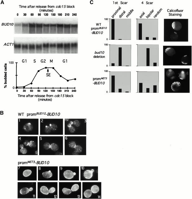
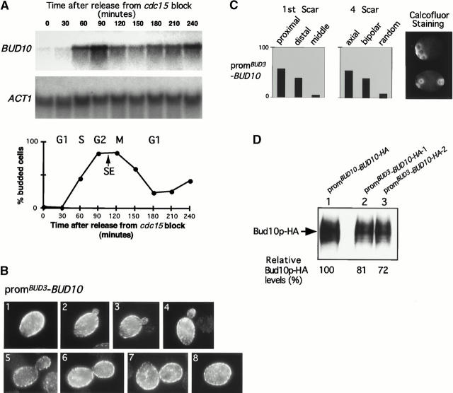
Genomic studies in yeast have revealed that one eighth of genes are cell cycle regulated in their expression. Almost without exception, the significance of cell cycle periodic gene expression has not been tested. Given that many such genes are critical to cellular morphogenesis, we wanted to examine the importance of periodic gene expression to this process. The expression profiles of two genes required for the axial pattern of cell division, BUD3 and BUD10/AXL2/SRO4, are strongly cell cycle regulated. BUD3 is expressed close to the onset of mitosis. BUD10 is expressed in late G1. Through promotor-swap experiments, the expression profile of each gene was altered and the consequences examined. We found that an S/G2 pulse of BUD3 expression controls the timing of Bud3p localization, but that this timing is not critical to Bud3p function. In contrast, a G1 pulse of BUD10 expression plays a direct role in Bud10p localization and function. Bud10p, a membrane protein, relies on the polarized secretory machinery specific to G1 to be delivered to its proper location. Such a secretion-based targeting mechanism for membrane proteins provides cells with flexibility in remodeling their architecture or evolving new forms.


Figures

Figure 1
Figure 1
Cell cycle expression profiles of BUD3 and BUD10. (A) Northern blot of BUD3 and BUD10 expression during the cell cycle of wild-type cells (JC1362). Synchronization of the cell cycle was achieved through use of a cdc15-2ts background (Benton et al. 1997). LEU2 is a cell cycle–constitutive control. (B) Cell cycle stages of the synchronized culture defined by the budding index (scored by counting unbudded versus budded cells; see Materials and Methods for details). SE denotes the initiation of spindle elongation. (C) Cell cycle stages of the synchronized culture were scored by microtubule morphologies.
Figure 3
Figure 3
Cell cycle–constitutive expression of BUD10. (A) Northern blot analysis of BUD10 expression in JC2133 carrying pJC1869 (promMET3-BUD10). The graph depicts the cell cycle stages of the synchronized culture as defined by the budding index and spindle elongation (SE). ACT1 is a cell cycle–constitutive control. (B) Localization of Bud10p during the cell cycle. The top images (1–6) show localization of HA-tagged Bud10p in haploids when expressed from its native promoter (JC1295 carrying pJC246 [promBUD10-BUD10]). The bottom images (7–14) show localization of HA-tagged Bud10p in JC1295 carrying pJC1869 (promMET3-BUD10). (C) Quantitation of the ability of the constitutively expressed BUD10 to support axial budding. Bud scar counts are shown for wild-type (JC1295 carrying pJC246), bud10-deletion (JC1295), and constitutive BUD10 (JC1295 carrying pJC1869) cells.
Figure 2
Figure 2
Cell cycle–constitutive expression of BUD3. (A) Northern blot analysis of BUD3 expression in JC2123 carrying pJC117 (promMET3-BUD3). The graph depicts the cell cycle stages of the synchronized culture as defined by the budding index and spindle elongation (SE). ACT1 is a cell cycle–constitutive control. (B) Localization of Bud3p during the cell cycle. The top images (1–4) show localization of Bud3p in strain JC1030 (anti-Bud3p antibody). The bottom images (5–8) show localization of HA-tagged Bud3p in JC1997 carrying pJC117 (promMET3-BUD3) under inducing conditions. (C) The ability of constitutively expressed BUD3 to support axial budding. Bud scar counts are shown for wild-type cells (JC1030), bud3-deletion cells (JC1997), and constitutive BUD3 cells (JC1997) carrying pJC117. For each, the arrangements of scars on cells with one bud scar and cells with four bud scars were scored relative to the birth scar after staining with calcofluor (Chant and Pringle 1995). (D) Examples of cells exhibiting defects in morphology and septin (Cdc3p) localization upon overexpression of BUD3. EJY301 carrying the promGAL1-BUD3 overexpression construct (pJC16) was grown in glucose-containing (repressing) or galactose-containing (inducing) media.
Figure 4
Figure 4
Expression of BUD10 from the BUD3 promoter. (A) Northern blot analysis of BUD10 expression in JC2133 carrying pJC256 (promBUD3-BUD10). The graph depicts the cell cycle stages of the synchronized culture as defined by the budding index and spindle elongation (SE). ACT1 is a cell cycle–constitutive control. (B) Localization of Bud10p during the cell cycle. The images (1–8) show the localization of HA-tagged Bud10p in JC1295 carrying pJC256 (promBUD3-BUD10). For comparison with the localization of Bud10p in wild-type cells, see Fig. 3 B. (C) Quantitation of the ability of the construct containing BUD10 under the control of the BUD3 promoter to support axial budding. Bud scar counts of JC1295 carrying pJC256 (promBUD3-BUD10). For comparison with wild-type and bud10 cells see Fig. 3 C. (D) Western blot measurement of Bud10-HAp levels in JC1295 carrying Bud10-HAp under its native promoter (pJC246, lane 1) or under the control of the BUD3 promoter (pJC256 and pJC257, lanes 2 and 3, respectively). Equal amounts of protein generated from cell extracts were loaded in each lane, and relative levels of Bud10-HAp were quantitated (see Materials and Methods).
Figure 5
Figure 5
Pulsing BUD10 expression in an asynchronous culture. (A) Quantitation of the distribution of HA-tagged Bud10p signal in JC1296 carrying pJC1869 (promMET3-BUD10). At each time point after pulsing of BUD10 expression from the MET3 promoter (0, 60, and 120 min), the distribution of Bud10p signal was placed into three categories: correctly localized, mislocalized, and intracellular. (B) Position in the cell cycle at which a correctly localized HA-tagged Bud10p signal was apparent by immunofluorescence analysis. (C) Immunofluorescence analysis of HA-tagged Bud10p after a brief induction of BUD10-HA expression. At each time point after induction, examples of correctly localized Bud10p (cells 1–4) and incorrectly localized Bud10p signals (cells 5–8) are shown.
Figure 6
Figure 6
Pulsing BUD10 expression in synchronous cultures. (A) The graph depicts synchrony profiles of JC2133 cultures carrying pJC1869 (promMET3-BUD10), which were pulsed for BUD10 expression. The cell cycle stage was defined by the budding index and spindle elongation (SE). Late G1 and S/G2 inductions of BUD10 expression were achieved by a 45-min incubation of cell samples in methionine-deficient medium for the periods indicated with double headed arrows. Under these conditions, the cell cycle length (cell doubling time) was ∼210 min. (B) The percent distribution of Bud10-HAp signal found in the secretory pathway, correctly localized or mislocalized, was scored after induction centered on late G1 phase. Examples of each type of localization are shown above the values: cell 1, secretory pathway localization; cells 2 and 3, correctly localized; and cell 4, mislocalized signal. (C) The percent distribution of Bud10-HAp signal found in the secretory pathway, correctly localized or mislocalized in the cell, was scored after induction centered on S/G2 phase. Examples of each type of localization, as detected by immunofluorescence, are shown above the values: cell 1, secretory pathway localization; cell 2, correctly localized; and cells 3 and 4, mislocalized signal.
Figure 7
Figure 7
Proposed localization mechanisms of Bud3p and Bud10p. Cell cycle expression of BUD3 leads to the production of cytoplasmic Bud3p (red), which localizes by diffusion and docking at the septin ring. BUD10 is expressed in G1 at which time secretion is tightly focused on the nascent bud site. Bud10p (green) is delivered to the bud site and remains for the duration of the cell cycle in this position, which becomes the mother–bud neck.

Similar articles

Cited by

References

    1. Bender A., Pringle J.R. Multicopy suppression of the cdc24 budding defect in yeast by CDC42 and three newly identified genes including the ras-related RSR1 . Proc. Natl. Acad. Sci. USA. 1989;86:9976–9980. - PMC - PubMed
    1. Benton B.K., Tinkelenberg A., Gonzalez I., Cross F.R. Cla4p, a Saccharomyces cerevisiae Cdc42p-activated kinase involved in cytokinesis, is activated at mitosis. Mol. Cell. Biol. 1997;17:5067–5076. - PMC - PubMed
    1. Byers B., Goetsch L. A highly ordered ring of membrane-associated filaments in budding yeast. J. Cell. Biol. 1976;69:717–721. - PMC - PubMed
    1. Carroll C.W., Altman R., Schieltz D., Yates J.R., Kellogg D. The septins are required for the mitosis-specific activation of the Gin4 kinase. J. Cell Biol. 1998;143:709–717. - PMC - PubMed
    1. Chant J., Herskowitz I. Genetic control of bud site selection in yeast by a set of gene products that constitute a morphogenetic pathway. Cell. 1991;65:1203–1212. - PubMed

Publication types

MeSH terms