. 2000 Dec 25;151(7):1583-90.
doi: 10.1083/jcb.151.7.1583.

Loss of calpain 3 proteolytic activity leads to muscular dystrophy and to apoptosis-associated IkappaBalpha/nuclear factor kappaB pathway perturbation in mice

Affiliations

Loss of calpain 3 proteolytic activity leads to muscular dystrophy and to apoptosis-associated IkappaBalpha/nuclear factor kappaB pathway perturbation in mice

I Richard et al. J Cell Biol. .

Abstract

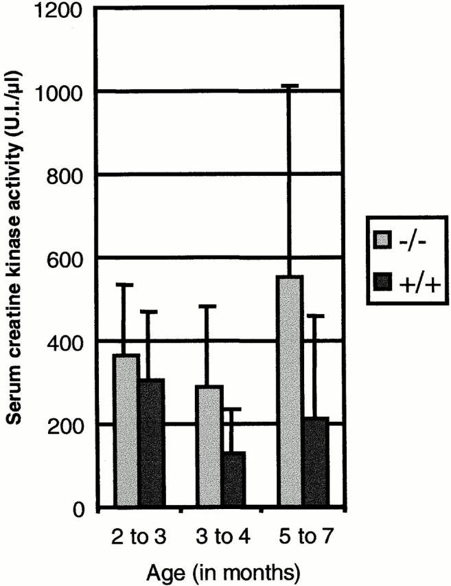
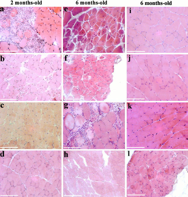
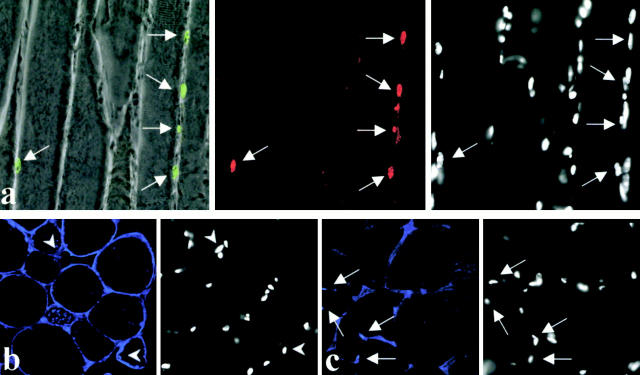
Calpain 3 is known as the skeletal muscle-specific member of the calpains, a family of intracellular nonlysosomal cysteine proteases. It was previously shown that defects in the human calpain 3 gene are responsible for limb girdle muscular dystrophy type 2A (LGMD2A), an inherited disease affecting predominantly the proximal limb muscles. To better understand the function of calpain 3 and the pathophysiological mechanisms of LGMD2A and also to develop an adequate model for therapy research, we generated capn3-deficient mice by gene targeting. capn3-deficient mice are fully fertile and viable. Allele transmission in intercross progeny demonstrated a statistically significant departure from Mendel's law. capn3-deficient mice show a mild progressive muscular dystrophy that affects a specific group of muscles. The age of appearance of myopathic features varies with the genetic background, suggesting the involvement of modifier genes. Affected muscles manifest a similar apoptosis-associated perturbation of the IkappaBalpha/nuclear factor kappaB pathway as seen in LGMD2A patients. In addition, Evans blue staining of muscle fibers reveals that the pathological process due to calpain 3 deficiency is associated with membrane alterations.


Figures

Figure 1
Figure 1
Gene targeting of mice. (a) Restriction map of the targeting construct, the wild-type capn3 locus, and the targeted locus. A fragment containing exons 2 and 3 encompassing C129, the cysteine participating to the catalytic site, was deleted and replaced by a neoR cassette. The herpes simplex virus tk (TK) cassette was added 5′ to the targeting vector for positive–negative selection. H, E, and K indicate HindIII, EcoRI, and KpnI, respectively. (b) Double-resistant ES cell clones were genotyped by long-range PCR using two primer pairs (5′.a/Neo.1 and Neo.2/3′.m). Primer sites are shown in panel a. The 6- and 2.1-kb PCR products are indicative of correct homologous recombination events in the 5′ and 3′ neoR flanking regions, respectively. (c) Genotype analysis of progeny from a heterozygote intercross. PCR analysis of tail DNA isolated from 3-wk-old mice was performed using the PCR primer pairs Neo.3/ex4.m (top) and ex3.a/ex3.m (bottom). Normal mice DNA (+/+) gave an amplification product only with exon 3 primer pairs; capn3 −/− mice DNA (−/−), only with neo primer pairs; and capn3 +/− mice DNA gave amplification products with both sets of primers. (d) Quantitative RT-PCR analysis of calpain 3 messenger level, expressed relative to wild-type mice, in RNA extracted from muscle. The analysis was performed according to TaqMan chemistry using primer pairs located in exon 1. Level of TFIID mRNA was used to normalize the results across different samples. The bars indicate the mean value ± the minimum or maximum. (e) Western blot analysis. A calpain 3–specific polyclonal antibody directed against the IS2 region of calpain 3 was used to detect calpain 3 expression in the muscle of wild-type (+/+) and capn3 −/− (−/−) mice. A Western blot using a calpain 2–specific polyclonal antibody was used as a control. Zinc staining was used to ascertain equivalent loading. (f) Western blot analyses were performed on lysates of transfected COS-7 cells to visualize proteolytic calpain 3 fragments using an antibody against the IS2-specific region of p94. The constructs used were the vector alone (pSRD) or derived constructs containing, respectively, the wild-type calpain 3 (p94), the cDNA corresponding to the calpain 3–deficient mice (KO), and a defective cDNA carrying the mutation C129S. White and black arrowheads indicate the full-length and proteolyzed products, respectively. (g) Proteolysis of fodrin was assessed using antibodies specific to the 150-kD α-fodrin fragment. The proteolized fragment was only detected for the wild-type calpain 3. (h) The titin-binding capacities of the cDNA was monitored in a yeast two-hybrid system, using as bait two different titin clones corresponding to the COOH-terminal (C-ter) region (pCNT-52) and to the region located in the N2A line of the sarcomere (pCNT-N2). Wild-type (p94), mutant calpain 3 (KO) cDNA, or vector alone (T-[pAS2C-1]) was cotransfected with the titin clones into Saccharomyces cerevisiae CG-1945 strain cells. Binding was visualized as growth on plates without leucine and without tryptophane. Deletion of exons 2 and 3 does not impair binding to the N2A region of titin but weakens binding to the COOH-terminal region.
Figure 2
Figure 2
Serum CK activity. This parameter was evaluated on the 129Sv background. Animals were grouped into three classes according to their age. For the 2–3-mo-old animals, n = 38 for −/−, n = 25 for +/+; for the 3–4-mo-old animals, n = 68 for −/−, n = 6 for the +/+; and for the 5–7-mo-old animals, n = 37 for −/− and n = 21 for +/+. The bars indicate the mean values ± SD. P < 0.09 at 2–3 mo of age, P < 0.03 at 3–5 mo of age, and P < 0.002 at 6 mo of age.
Figure 3
Figure 3
Histologic analysis of capn3-deficient mice. H&E-stained transverse muscle cryosections (7 μm) from mice of 129Sv genetic background. (a–d) 2-mo-old mice; (e–l) 6-mo-old mice. Dystrophic changes are evident in psoas (a and e), soleus (b and f), deltoid (c and g), and tibialis anterior (d and h), whereas quadriceps (i), gastrocnemius (j), and triceps (k) present a normal aspect. Diaphragm in l presents very slight abnormalities. The main features encountered are fibers with internal nuclei (b–f, and h) or area of infiltration of mononuclear cells (a and g). A cluster of small regenerating fibers can be seen in e. Bars, 50 μm.
Figure 4
Figure 4
Apoptosis and immunolocalization of IκBα and NF-κB in muscles of calpain 3–deficient mice. (a) A longitudinal muscle section of a calpain 3–deficient mouse was triple labeled for apoptosis (green myonuclei superimposed over the phase–contrast field), IκBα (red), and chromatin (DAPI, right). Note that TUNEL-positive myonuclei were always IκBα-positive (arrowheads). (b) Subsarcolemmal localization of NF-κB (blue fluorescence, left). Note that in this calpain 3–deficient tissue, NF-κB was not found concentrated in the myonuclei (stained with DAPI, right, arrowheads). (c) As a control, NF-κB (blue fluorescence) was normally found concentrated in some myonuclei of wild-type mice (arrowheads). The DAPI fields were recorded with a CCD camera mounted on an additional port of the confocal microscope. For a, 10 mm corresponds to 72.3 μm, and for b and c, 10 mm corresponds to 57 μm.
Figure 5
Figure 5
EB staining of cryosections of calpain 3–deficient muscles reveals apoptosis-associated disruption of sarcolemmal integrity. Four 6-mo-old animals of each genotype were killed 12 h after being injected with EB dye and biopsied. Regions of EB uptake are seen as red cytoplasmic staining on fluorescence microscopy. A mean of three EB-positive cells was detected per muscle section of calpain 3–deficient mice. (a) Calpain 3–deficient biceps showing EB staining. No EB uptake is seen in wild-type biceps. (b) Triple labeling for EB, TUNEL, and propidium iodide (PI) of a muscle section of calpain 3–deficient mouse. From left to right, EB-positive cells (red pseudocolor), TUNEL-positive myonuclei, propidium iodide–stained nuclei (blue pseudocolor), and a superposition of all stainings (white arrows and/or green pseudocolor refer to apoptotic nuclei). Bars: (a) 50 μm; (b) 20 μm.

Similar articles

Cited by

References

    1. Baghdiguian S., Martin M., Richard I., Pons F., Astier C., Bourg N., Hay R.T., Chemaly R., Halaby G., Loiselet J. Calpain 3 deficiency is associated with myonuclear apoptosis and profound perturbation of the IκBa/NF-kB pathway in limb-girdle muscular dystrophy type 2A. Nat. Med. 1999;5:503–511. - PubMed
    1. Baldwin A.S., Jr. The NFκB and IκB proteinsnew discoveries and insights. Annu. Rev. Immunol. 1996;14:649–681. - PubMed
    1. Bönnemann C.G., Modi R., Noguchi S., Mizuno Y., Yoshida M., Gussoni E., McNally E.M., Duggan D.J., Angelini C., Hoffman E.P., Ozawa E., Kunkel L.M. β-Sarcoglycan (A3b) mutations cause autosomal recessive muscular dystrophy with loss of the sarcoglycan complex. Nat. Genet. 1995;11:266–273. - PubMed
    1. Dear T.N., Boehm T. Diverse mRNA expression patterns of the mouse calpain genes Capn5, Capn6 and Capn11 during development. Mech. Dev. 1999;89:201–209. - PubMed
    1. Dourmashkin R.R., Deteix P., Simone C.B., Henkart P. Electron microscopic demonstration of lesions in target cell membranes associated with antibody-dependent cellular cytotoxicity. Clin. Exp. Immunol. 1980;42:554–560. - PMC - PubMed

Publication types

MeSH terms