. 2001 Jan;107(1):83-90.
doi: 10.1172/JCI10224.

An etiological role for aeroallergens and eosinophils in experimental esophagitis

Affiliations

An etiological role for aeroallergens and eosinophils in experimental esophagitis

A Mishra et al. J Clin Invest. 2001 Jan.

Abstract

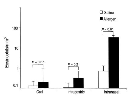
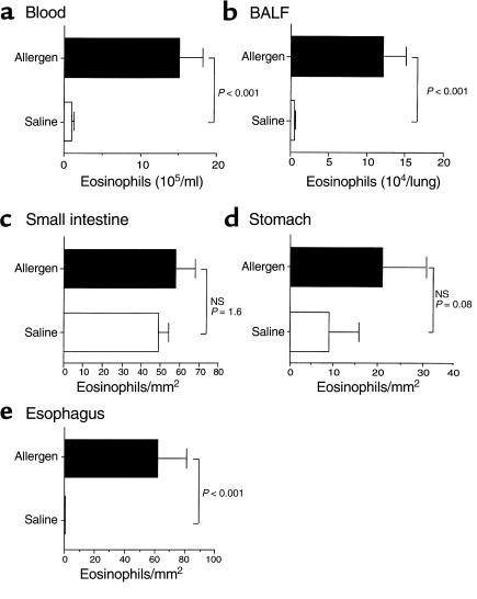
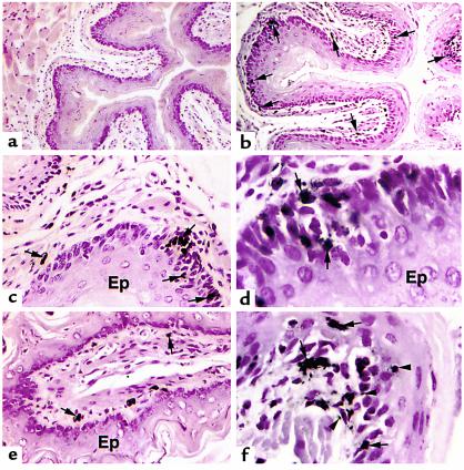
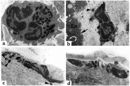
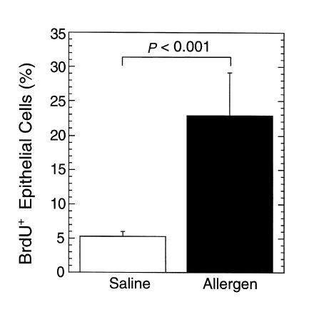
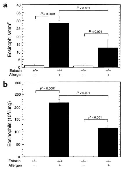
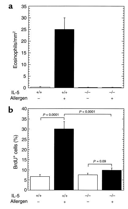
Eosinophil infiltration into the esophagus is observed in diverse diseases including gastroesophageal reflux and allergic gastroenteritis, but the processes involved are largely unknown. We now report an original model of experimental esophagitis induced by exposure of mice to respiratory allergen. Allergen-challenged mice develop marked levels of esophageal eosinophils, free eosinophil granules, and epithelial cell hyperplasia, features that mimic the human disorders. Interestingly, exposure of mice to oral or intragastric allergen does not promote eosinophilic esophagitis, indicating that hypersensitivity in the esophagus occurs with simultaneous development of pulmonary inflammation. Furthermore, in the absence of eotaxin, eosinophil recruitment is attenuated, whereas in the absence of IL-5, eosinophil accumulation and epithelial hyperplasia are ablated. These results establish a pathophysiological connection between allergic hypersensitivity responses in the lung and esophagus and demonstrate an etiologic role for inhaled allergens and eosinophils in gastrointestinal inflammation.


Figures

Figure 1
Figure 1
The effect of oral, intragastric, and intranasal allergen challenge on eosinophil levels in the esophagus. Mice (BALB/c) were challenged with repeated inoculations of oral, intragastric and intranasal allergen. Eighteen hours after the last allergen or placebo challenge, the mice were sacrificed and eosinophil levels were evaluated in the esophagus. Data are expressed as mean ± SEM; n = 8–9 mice.
Figure 2
Figure 2
The effect of intranasal allergen challenge on eosinophil levels. Mice were challenged with allergen or saline intranasally, and 18 hours after the last allergen exposure the mice were sacrificed and eosinophil levels in the blood (a), BALF (b), small intestine (c), stomach (d), and esophagus (e) were determined. Data are expressed as mean ± SEM (n = 8–12 mice in each group). The mouse strains were a mixture of 129 SvEv, BALB/c, and FVB/N. NS, not significant.
Figure 3
Figure 3
Light microscopic analysis of eosinophils in the esophagus. Immunohistochemical identification (by anti-MBP staining) of eosinophils in the esophagus of saline-challenged (a) and allergen-challenged (bf) wild-type (BALB/c) mice. Most eosinophils (arrows) are located in the lamina propria (bf), submucosa (e), and epithelial layer (d and f). Higher-power magnification photomicrographs reveal penetration of eosinophils through the basal epithelial layer (d) and the detection of extracellular MBP staining (f; arrowheads). Original magnifications are ×150 (a), ×250 (b), ×500 (c and e), and ×1250 (d and f). Ep, epithelium.
Figure 4
Figure 4
Electron microscopic analysis of esophageal eosinophils. Mice (BALB/c) were treated with saline (a) or allergen (bd), and electron microscopy was performed on the esophagus. Representative electron micrographs are shown. (a) A healthy-appearing eosinophil with an intact plasma membrane, a segmented nucleus, and abundant electron-dense granules with characteristic cores and matrices. (bd) Eosinophil plasma membranes are disrupted, the nuclei are undergoing chromolysis, and there are readily detectable extracellular eosinophil granules (shown with arrowheads). The location of an eosinophil next to the epithelial layer (Ep) and below the basement membrane (arrow) is illustrated in b. Original magnifications are ×8000 (a), ×5000 (b and d), and ×6300 (d).
Figure 5
Figure 5
Epithelial proliferation following allergen challenge. Mice (BALB/c) were challenged with saline or allergen intranasally and the incorporation of BrdU in the epithelial layer was measured 3 hours after the last challenge. The results are mean ± SEM (n = 4–5 mice).
Figure 6
Figure 6
Induction of eosinophilic esophagitis in eotaxin gene-targeted mice. Wild-type (+/+) or eotaxin-deficient (–/–) mice (BALB/c) were challenged with control saline (–) or allergen (+), and the number of eosinophils in the esophagus (a) was determined by anti-MBP staining and in the lung fluid (b) by differential counting of cells in BALF. The results are expressed as mean ± SEM (n = 4–6 mice).
Figure 7
Figure 7
The role of IL-5 in allergen-induced eosinophil recruitment and epithelial hyperplasia in the esophagus. Wild-type (+/+) and IL-5 gene-targeted (–/–) mice (C57BL/6) were subjected to the allergen challenge protocol by treatment with control saline (–) or allergen (+) inoculations. (a) The number of eosinophils in the esophagus was determined by anti-MBP staining. (b) The incorporation of BrdU in the epithelial layer was measured 3 hours after the last challenge. The results are mean ± SEM (n = 4–5 mice).

References

    1. Holgate ST. The epidemic of allergy and asthma. Nature. 1999;402:B2–B4. - PubMed
    1. Sampson HA. Food allergy. Part 1: immunopathogenesis and clinical disorders. J Allergy Clin Immunol. 1999;103:717–728. - PubMed
    1. Furuta GT, Ackerman SJ, Wershil BK. The role of the eosinophil in gastrointestinal diseases. Curr Opin Gastroenterol. 1995;11:541–547.
    1. Burks AW. The spectrum of food hypersensitivity: where does it end? J Pediatr. 1998;133:175–176. - PubMed
    1. Winter HS, et al. Intraepithelial eosinophils: a new diagnostic criterion for reflux esophagitis. Gastroenterology. 1982;83:818–823. - PubMed

Publication types

MeSH terms