. 2001 Jan;107(1):99-109.
doi: 10.1172/JCI10501.

Salmonella typhimurium translocates flagellin across intestinal epithelia, inducing a proinflammatory response

Affiliations

Salmonella typhimurium translocates flagellin across intestinal epithelia, inducing a proinflammatory response

A T Gewirtz et al. J Clin Invest. 2001 Jan.

Abstract

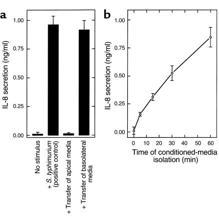
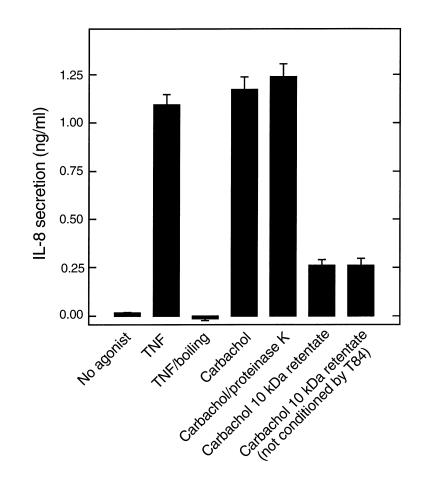
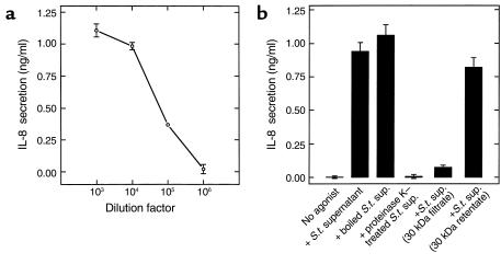
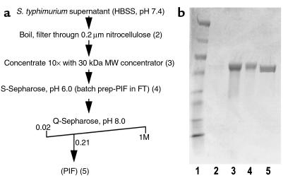
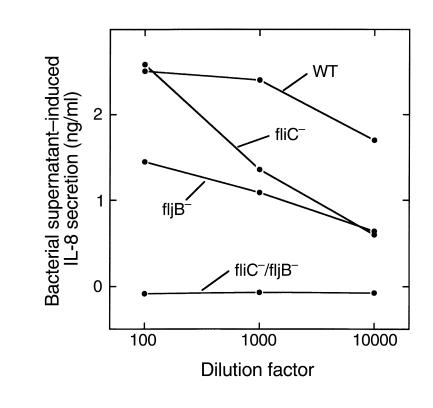
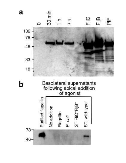
This study investigated whether soluble paracrine factors mediated Salmonella-induced IL-8 expression in polarized model intestinal epithelia. We found that the basolateral media of model epithelia that had been apically infected with Salmonella typhimurium for a short period (10 minutes) could activate IL-8 secretion in virgin model epithelia, demonstrating that a proinflammatory factor (PIF) was indeed present. Initial characterization found that PIF was a heat-stable protein with a molecular mass of about 50 kDa that acts on the basolateral, but not apical, surface of model intestinal epithelia to elicit IL-8 secretion. PIF was not present in the media of model epithelia stimulated with other inducers of IL-8 secretion (TNF-alpha or carbachol) but was present in S. typhimurium supernatants, indicating PIF is of bacterial origin. PIF was purified from bacterial culture supernatants by anion/cation exchange chromatography and SDS-PAGE and found by using microsequencing to be the protein flagellin. In support of this finding, flagellin-deficient S. typhimurium mutants did not secrete detectable levels of PIF (i.e., a bioactivity that induced IL-8 secretion when placed basolaterally on model epithelia). Furthermore, viable flagellin-deficient mutant organisms (fliC/fljB and flhD) failed to elicit IL-8 secretion when added apically to model intestinal epithelia. These findings indicate that translocation of flagellin across epithelia, subsequent to apical epithelial-S. typhimurium interaction, is likely a major means of activating a mucosal inflammatory response.


Figures

Figure 1
Figure 1
Basolaterally conditioned media of S. typhimurium–infected epithelia induce IL-8 secretion in virgin epithelia. Model intestinal epithelia were apically exposed to S. typhimurium. The epithelial media was then transferred to untreated model epithelia. IL-8 was assayed in basolateral media 5 hours later. (a) Media was transferred maintaining its polarity of origin (i.e., apical to apical, basolateral to basolateral) 1 hour after addition of bacteria. Live S. typhimurium (added apically for 5 hours) served as a positive control. (b) Basolateral media was transferred at the indicated time after addition of bacteria.
Figure 2
Figure 2
PIF is a heat-stable protein. Samples from a and b were applied basolaterally to model epithelia, and their ability to induce IL-8 secretion over a 5-hour period was measured. (a) PIF was centrifuged through indicated Amicon concentrators followed by dilution of retentate to original volume. (b) PIF, TNF-α (20 ng/ml), and carbachol (100 μM) were subjected to boiling (20 minutes) or proteinase K treatment (1 mg/ml for 1 hour followed by 10 minutes of boiling to inactivate proteinase K).
Figure 3
Figure 3
Epithelia do not release PIF in response to TNF-α or carbachol. Model epithelia were basolaterally treated with TNF-α (20 ng/ml) or carbachol (100 μM). One hour later, basolateral media (which still contained TNF-α or carbachol) was isolated and subjected to boiling or proteinase K treatment where indicated (as described in Figure 2). Additionally, carbachol-containing media (and an equimolar solution of carbachol that had not been exposed to epithelia) were concentrated tenfold over a 10-kDa Amicon concentrator and then rediluted to the original volume. Samples were then basolaterally exposed to fresh model epithelia, and their ability to induce IL-8 secretion over a 5-hour period was measured.
Figure 4
Figure 4
Bacteria release a PIF with the same properties as that isolated from bacterial-epithelial interactions. An overnight culture of S. typhimurium (S.t.) was pelleted, washed two times by centrifugation, and resuspended in HBSS (1010 CFU/ml). One hour later, bacterial supernatant was isolated and basolaterally applied to model epithelia where its ability to induce IL-8 secretion was measured.(a) Supernatant was applied over a range of concentrations. (b) Bacterial supernatant was subjected to indicated treatments (as described in Figure 2) before being applied to epithelia.
Figure 5
Figure 5
Purification of PIF revealed it to be the protein flagellin. (a) Outline of PIF purification method. Numbers in parentheses correspond to lane numbers in b. (b) Coomassie-stained gel showing PIF at various stages of purification. Lane 1: MW markers (in kDa: 205, 120, 84, 52, 36, 30, 22, 7). Lane 2: S. typhimurium supernatant (i.e., starting material; too dilute to detect any bands). Lane 3: Sample concentrated tenfold over 30-kDa cut-off Amicon filter. Lane 4: Concentrated sample after incubation with S-Sepharose cation exchange beads. Lane 5: The peak bioactive anion exchange fraction. This band was sequenced and found to be flagellin. FT, flow through.
Figure 6
Figure 6
Flagellin-deficient S. typhimurium do not release an IL-8 inducing bioactivity. Bacterial supernatants were isolated (as described in Figure 4) from wild-type (WT) S. typhimurium or mutant strains lacking fliC and/or fljB (“–” indicates the gene that is missing). Supernatants were then diluted as indicated, applied basolaterally to model epithelia, and IL-8 secretion assayed 5 hours later.
Figure 7
Figure 7
Various bacteria release PIF/flagellin. An overnight culture of the indicated bacterial strain was pelleted, washed two times by centrifugation, and resuspended in HBSS (1010 CFU/ml). One hour later, bacterial supernatant was isolated. Flagellin-deficient strain (fliC/fljB) transformed with plasmids encoding fliC and fljB is indicated by fliC/fljB/fliC+ and fliC/fljB/fljB+, respectively. (a) Supernatants were analyzed by SDS-page/immunoblotting using a mAb to flagellin. (bd) Model epithelia were placed in HBSS. (b) Bacterial supernatants were applied basolaterally at a 1:100 dilution to model epithelia. IL-8 secretion was assayed 5 hours later. (c) Bacterial supernatants were applied basolaterally, at indicated dilution, to model epithelia. IL-8 secretion was assayed 5 hours later. (d) Live bacteria were applied to apical reservoir (108 bacteria/ epithelia). IL-8 secretion was assayed 5 hours later.
Figure 8
Figure 8
Basolateral flagellin, but not apical flagellin, induces IL-8 secretion. Flagellin was isolated from S. typhimurium supernatant as described in Methods, diluted as indicated, and added to the indicated reservoir of model epithelia, and IL-8 secretion was measured 5 hours later.
Figure 9
Figure 9
Flagellin rapidly appears in the basolateral supernatants of apically infected model epithelia. Model intestinal epithelia were apically exposed to indicated bacteria. Basolateral epithelial supernatants were isolated and Western blotted for flagellin. (a) Supernatants were isolated at the indicated time after addition of S. typhimurium. Isolated surface flagellin from S. typhimurium expressing FliC or FljB and PIF were directly Western blotted (i.e., not exposed to epithelia) to serve as positive controls. (b) Supernatants were isolated 1 hour after exposure to indicated organism or purified flagellin (100 ng/ml).
Figure 10
Figure 10
Flagellin translocation by S. typhimurium mutants. Model epithelia were apically exposed to wild-type (WT) S. typhimurium or indicated mutant or nonpathogenic E. coli. for 45 minutes. After exposure to bacteria, basolateral supernatants were collected, boiled, filtered (through 2-μM pore size filter), and transferred to basolateral surface of virgin epithelia. IL-8 secretion was measured 5 hours later using ELISA. Wild-type represents PhoPc parent (14028s). An indistinguishable result was obtained using invA parent SR-11 (not shown).
Figure 11
Figure 11
Flagellin is a potent inducer of IL-8 secretion, does not drop transepithelial electrical resistance (TER), and is essential for IL-8 secretion induced by S. typhimurium. (a) Model epithelia were basolaterally exposed to indicated concentration of LPS, flagellin, or TNF-α. IL-8 secretion was assayed 5 hours later. (b) Model epithelia were exposed (basolaterally) to HBSS (Neg), PIF (1:40 of sample used in Figure 4), TNF-α (20 ng/ml), or C. difficile toxin A (Toxin A). TER was measured as described in Methods. (c) Model epithelia were apically colonized for 45 minutes with live indicated wild-type (WT) S. typhimurium or indicated mutant strain. Nonadherent bacteria were removed by washing three times with HBSS. IL-8 secretion was measured 5 hours after initial exposure to bacteria. Flagellin-deficient strain (fliC/fljB) transformed with plasmids encoding fliC and fljB is indicated by fliC/fljB/fliC+ and fliC/fljB/fljB+, respectively.

Comment in

References

    1. Takeuchi A. Electron microscope studies of experimental Salmonella infection. Am J Pathol. 1967;50:109–119. - PMC - PubMed
    1. Madara JL. Review article: pathobiology of neutrophil interactions with intestinal epithelia. Aliment Pharmacol Ther. 1997;11(Suppl. 3):57–62. - PubMed
    1. McCormick BA, Parkos CA, Colgan SP, Carnes DK, Madara JL. Apical secretion of a pathogen-elicited epithelial chemoattractant (PEEC) activity in response to surface colonization of intestinal epithelia by Salmonella typhimurium. J Immunol. 1998;160:455–456. - PubMed
    1. McCormick BA, Colgan SP, Archer CD, Miller SI, Madara JL. Salmonella typhimurium attachment to human intestinal epithelial monolayers: transcellular signalling to subepithelial neutrophils. J Cell Biol. 1993;123:895–907. - PMC - PubMed
    1. Jung HC, et al. A distinct array of proinflammatory cytokines is expressed in human colon epithelial cells in response to bacterial invasion. J Clin Invest. 1995;95:55–65. - PMC - PubMed

Publication types