. 2001 Jan;75(2):952-60.
doi: 10.1128/JVI.75.2.952-960.2001.

Coxsackievirus A9 VP1 mutants with enhanced or hindered A particle formation and decreased infectivity

Affiliations

Coxsackievirus A9 VP1 mutants with enhanced or hindered A particle formation and decreased infectivity

A Airaksinen et al. J Virol. 2001 Jan.

Abstract

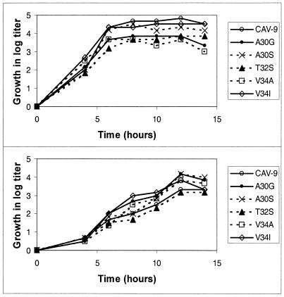
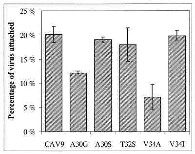
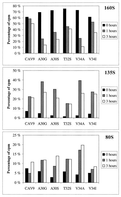
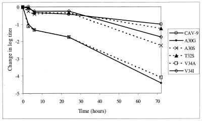
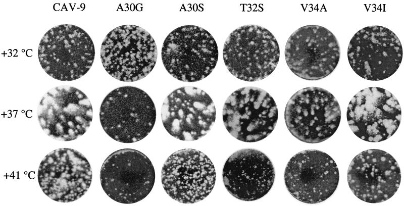
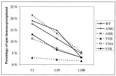
We have studied coxsackievirus A9 (CAV9) mutants that each have a single amino acid substitution in the conserved 29-PALTAVETGHT-39 motif of VP1 and a reduced capacity to produce infectious progeny virus. After uncoating, all steps in the infection cycle occurred according to the same kinetics as and similar efficiency to the wild-type virus. However, the particle/infectious unit ratio in the progeny was significantly increased. The differences were apparently due to altered stability of the capsid: there were mutant viruses with enhanced or hindered uncoating, and both of these characteristics were found to reduce fitness under standard passaging conditions. At 32 degrees C the instable mutants had an advantage, while the wild-type and the most stable mutant grew poorly. When comparing the newly published CAV9 structure and the other enterovirus structures, we found that the PALTAVETGHT motif is always in exactly the same position, in a cavity formed by the 3 other capsid proteins, with the C terminus of VP4 between this motif and the RNA. In the 7 enterovirus structures determined to date, the most conserved residues of the studied motif have identical contacts to neighboring residues of VP2, VP3, and VP4. We conclude that (i) the mutations affect the uncoating step necessary for infection, resulting in an untimely or hindered externalization of the VP1 N terminus together with the VP4, and (ii) the reason for the studied motif being evolutionarily conserved is its role in maintaining an optimal balance between the protective stability and the functional flexibility of the capsid.


Figures

FIG. 1
FIG. 1
Kinetics of production of infectious progeny virus at 37°C (top) and 32°C (bottom). To clarify the picture, the growth curves are shown as increase in titers after the attachment and washing steps.
FIG. 2
FIG. 2
Efficiency of attachment of CAV9 mutants to LLC Mk2 cells. The results shown are averages from four parallel experiments, and error bars indicate standard deviations.
FIG. 3
FIG. 3
Sedimentation analysis of CAV9 uncoating after incubating cell-bound virus for 0, 1, or 3 h at 37°C. Prior to this incubation, 35S-labeled viruses (about 2.5 × 10⁶ cpm) were allowed to adsorb to cells for 2 h at 18°C. Percentages of total label forming each peak (160S, 135S, and 80S) are shown.
FIG. 4
FIG. 4
Sedimentation analysis of spontaneous formation of subviral particles in medium. Percentages of total label as in the legend to Fig. 3 are shown.
FIG. 5
FIG. 5
Decrease of viral titers after incubation of 3, 6, 24, or 72 h in regular medium at 37°C. The results shown are averages of four parallel dilution series, comprising a total of 24 cell wells per dilution.
FIG. 6
FIG. 6
Plaque phenotypes at various temperatures.
FIG. 7
FIG. 7
Immunoprecipitation of CAV9 and mutants using three dilutions of peptide antiserum 910.
FIG. 8
FIG. 8
(A) Location of the VP1 hook in the three-dimensional structure of one protomer of CAV9, seen from the inside of the capsid. The tip of the hook dives in a cavity, the base of which is formed by the capsid proteins VP2 and VP3. The N termini of VP1, VP3, and VP4 are extended toward the fivefold axis at the bottom right. VP1, VP2, and VP3 are shown as space-filling models, while VP4 is shown as backbone only. The PALTAVETGHT motif is shown in cyan, while the rest of VP1 is blue. VP2, VP3, and VP4 are shown in yellow, red, and green, respectively. (B) A close view of the PALTAVETGHT motif and its surroundings in CAV9. Intraprotomer contact points that are completely conserved among PV1, PV2, PV3, CAV9, CBV5, echovirus 1, and BEV are shown as ball-and-stick models with dotted Van der Waals surfaces. The intertwined connection between the conserved VP3 amino acids 161 to 163 and VP1 amino acids 31 to 35 may have an important stabilizing role. Yellow text and arrows indicate the conserved amino acids, and white text and arrows indicate the amino acid chains included in this figure. Alpha carbons of conserved residues are colored white. CAV9 coordinates were from the Protein Data Bank of the Research Collaboratory for Structural Bioinformatics (available at http://www.rcsb.org/pdb/).

References

    1. Airaksinen A, Hovi T. Modified base compositions at degenerate positions of a mutagenic oligonucleotide enhance randomness in site-saturation mutagenesis. Nucleic Acids Res. 1998;26:576–581. - PMC - PubMed
    1. Airaksinen A, Roivainen M, Stanway G, Hovi T. Site-saturation mutagenesis of the PALTAVETG motif in coxsackievirus A9 capsid protein VP1 reveals evidence of conservation of a periodic hydrophobicity profile. J Gen Virol. 1999;80:1919–1927. - PubMed
    1. Arnold E, Rossmann M G. The use of molecular-replacement phases for the refinement of the human rhinovirus 14 structure. Acta Crystallogr A. 1988;44:270–282. - PubMed
    1. Basavappa R, Syed R, Flore O, Icenogle J P, Filman D J, Hogle J M. Role and mechanism of the maturation cleavage of VP0 in poliovirus assembly: structure of the empty capsid assembly intermediate at 2.9 Å resolution. Protein Sci. 1994;3:1651–1669. - PMC - PubMed
    1. Belnap D M, Filman D J, Trus B L, Cheng N, Booy F P, Conway J F, Curry S, Hiremath C N, Tsang S K, Steven A C, Hogle J M. Molecular tectonic model of virus structural transitions: the putative cell entry states of poliovirus. J Virol. 2000;74:1342–1354. - PMC - PubMed

Publication types

LinkOut - more resources