. 2001 Jan;75(2):1013-30.
doi: 10.1128/JVI.75.2.1013-1030.2001.

Accumulation of herpes simplex virus type 1 early and leaky-late proteins correlates with apoptosis prevention in infected human HEp-2 cells

Affiliations

Accumulation of herpes simplex virus type 1 early and leaky-late proteins correlates with apoptosis prevention in infected human HEp-2 cells

M Aubert et al. J Virol. 2001 Jan.

Abstract

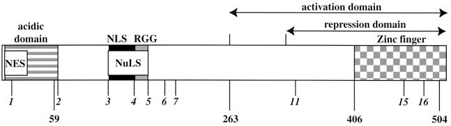
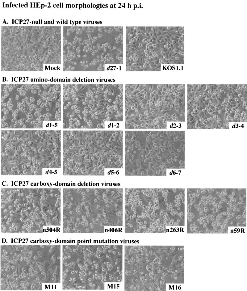
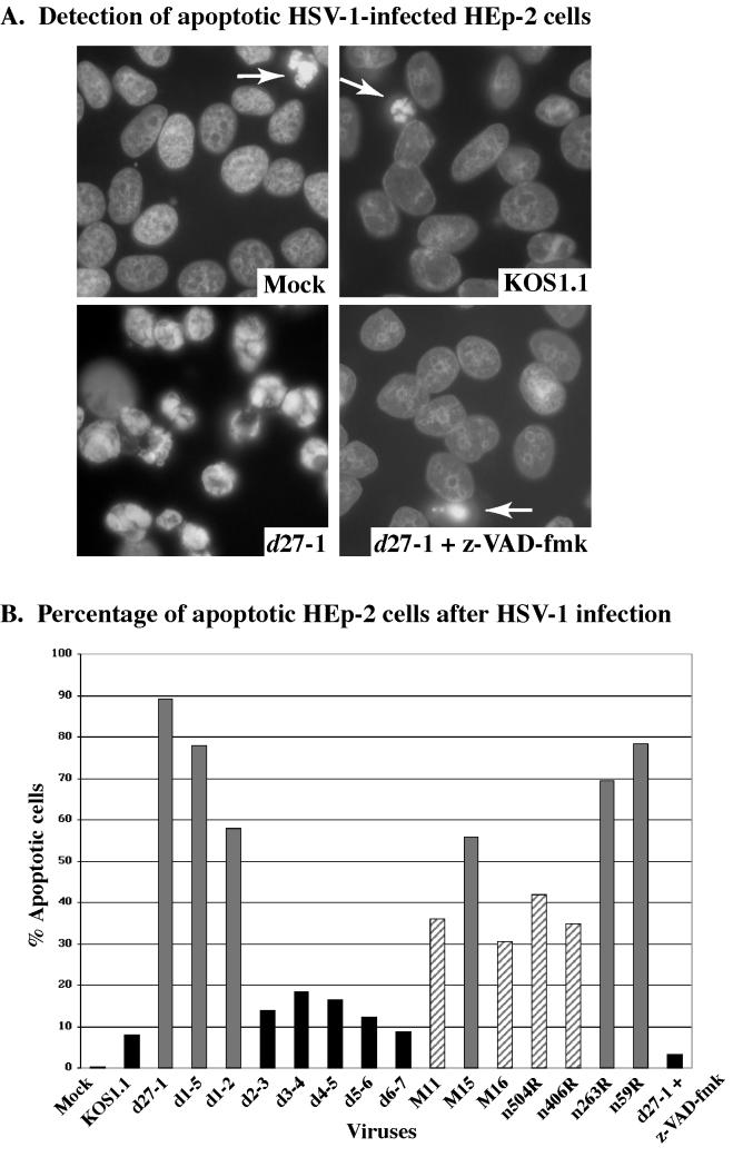
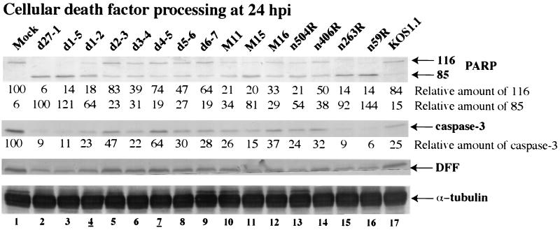
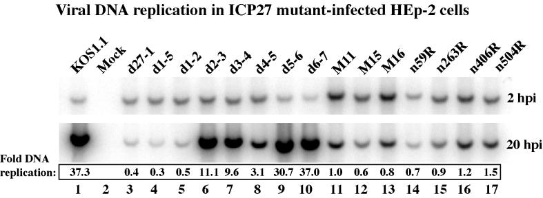
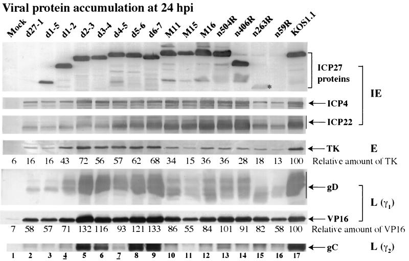
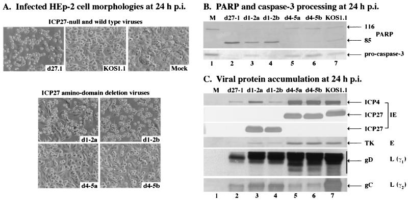
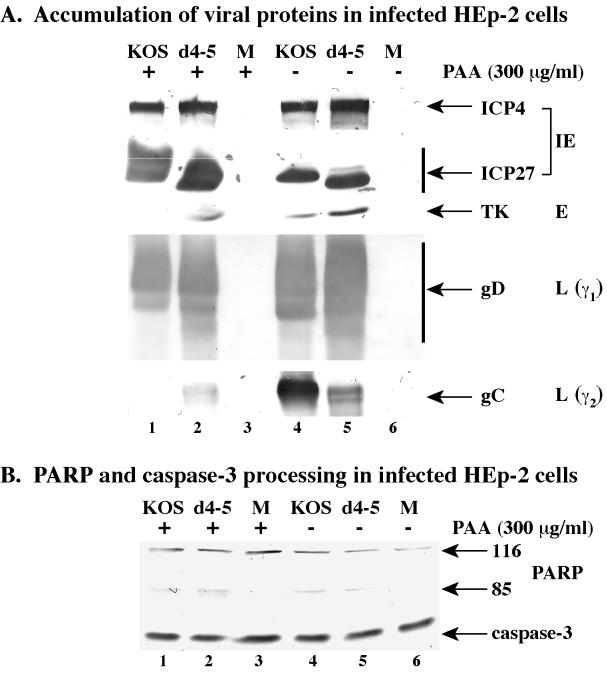
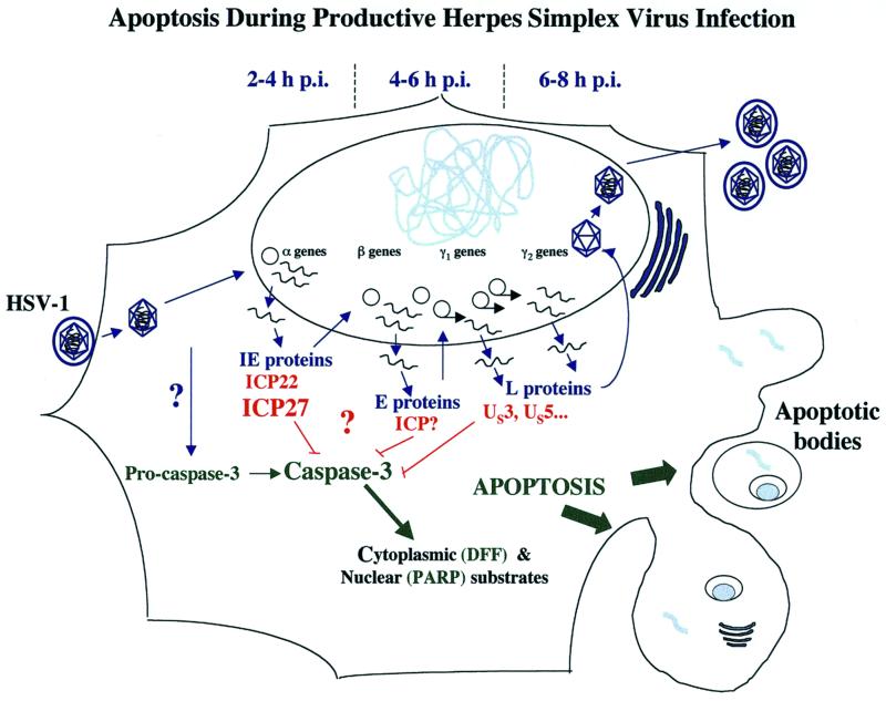
We previously reported that a recombinant ICP27-null virus stimulated, but did not prevent, apoptosis in human HEp-2 cells during infection (M. Aubert and J. A. Blaho, J. Virol. 73:2803-2813, 1999). In the present study, we used a panel of 15 recombinant ICP27 mutant viruses to determine which features of herpes simplex virus type 1 (HSV-1) replication are required for the apoptosis-inhibitory activity. Each virus was defined experimentally as either apoptotic, partially apoptotic, or nonapoptotic based on infected HEp-2 cell morphologies, percentages of infected cells with condensed chromatin, and patterns of specific cellular death factor processing. Viruses d27-1, d1-5, d1-2, M11, M15, M16, n504R, n406R, n263R, and n59R are apoptotic or partially apoptotic in HEp-2 cells and severely defective for growth in Vero cells. Viruses d2-3, d3-4, d4-5, d5-6, and d6-7 are nonapoptotic, demonstrating that ICP27 contains a large amino-terminal region, including its RGG box RNA binding domain, which is not essential for apoptosis prevention. Accumulations of viral TK, VP16, and gD but not gC, ICP22, or ICP4 proteins correlated with prevention of apoptosis during the replication of these viruses. Of the nonapoptotic viruses, d4-5 did not produce gC, indicating that accumulation of true late gene products is not necessary for the prevention process. Analyses of viral DNA synthesis in HEp-2 cells indicated that apoptosis prevention by HSV-1 requires that the infection proceeds to the stage in which viral DNA replication takes place. Infections performed in the presence of the drug phosphonoacetic acid confirmed that the process of viral DNA synthesis and the accumulation of true late (gamma(2)) proteins are not required for apoptosis prevention. Based on our results, we conclude that the accumulation of HSV-1 early (beta) and leaky-late (gamma(1)) proteins correlates with the prevention of apoptosis in infected HEp-2 cells.


Figures

FIG. 1
FIG. 1
Schematic of the ICP27 protein showing locations of defined functional regions. These include NES, a putative nuclear export signal (open box); the acidic domain, involved in gene expression and DNA synthesis (striped box); NLS, nuclear localization signal (black box); NuLS, nucleolar localization signal (white box); RGG sequence, RNA binding motif (gray box); and activation and repression domains (arrows), which include a cysteine-histidine zinc-finger-like domain (checkered box). Italicized numbers define mutated sites (M11, M15, and M16) and the boundaries of in-frame deletions. Bold numbers correspond to relevant amino acid positions.
FIG. 2
FIG. 2
Morphologies of HEp-2 cells infected with all viruses used in this study. Cells infected with d27-1, KOS1.1, and mock-infected virus (A) or viruses infected with ICP27 with an amino domain deletion (B), ICP27 with a carboxy domain deletion (C), and ICP27 with a carboxy domain point mutation (D) were observed at 24 h p.i. by phase-contrast microscopy (magnification, ×20) as described in Materials and Methods.
FIG. 3
FIG. 3
Visualization (A) and quantitation (B) of apoptotic HSV-1-infected HEp-2 cells. Mock- and HSV-1-infected HEp-2 cells were fixed at 15 h p.i., stained with Hoechst 33258, and visualized by fluorescence microscopy (magnification, ×60) as described in Materials and Methods. Panel A shows representative images of stained nuclei. d27-1-infected cells show classic condensed chromatin. The few apoptotic cells detected in mock-, KOS1.1-, and d27-1-infected cells in the presence of the general caspase inhibitory peptide (z-VAD-fmk) are marked by arrows. The percentage of apoptotic cells was determined by counting the number of infected cells with condensed chromatin. Gray, striped, and black histograms refer to apoptotic, partially apoptotic, and nonapoptotic viruses, respectively.
FIG. 4
FIG. 4
Immunoblot detection of cellular death factor processing in infected HEp-2 cells. Whole-cell extracts prepared at 24 h p.i. were used for immunoblot analyses with anti-PARP, anti-DFF, anti-caspase-3, and anti-α tubulin antibodies. Amounts of full-length PARP, processed PARP, and caspase-3 were calculated relative to the amount of α tubulin as described in Materials and Methods. 116 and 85 refer to full-length and processed PARP, respectively. Lanes 4 and 7 are underlined to mark viruses analyzed in Fig. 7.
FIG. 5
FIG. 5
Autoradiographic images of viral DNA. Total DNA was isolated from infected HEp-2 cells at 2 and 20 h p.i., digested with PstI, separated in an agarose gel, transferred to a nylon membrane, and hybridized with a radioactive probe derived from a portion of the HSV-1 UL44 gene, prior to autoradiography, as described in Materials and Methods. Amounts of DNA were quantitated using a phosphorimager, and values of fold-DNA replication were calculated by dividing the radioactive signal at 20 h p.i. (replicated DNA) by the signal at 2 h p.i. (input DNA).
FIG. 6
FIG. 6
Immunoblot detection of viral protein accumulation in infected HEp-2 cells. Whole-cell extracts prepared at 24 h p.i. were used for immunoblot analyses with anti-ICP27, anti-ICP4, and anti-ICP22 (IE proteins), anti-TK (E protein), anti-VP16 and anti-gD (L proteins, γ1), and anti-gC (L protein, γ2) antibodies. Relative amounts of TK and VP16 were normalized to the amount of α tubulin and calculated as described in Materials and Methods. An asterisk marks n263R ICP27 protein since its accumulation is at low levels. Lanes 4 and 7 are underlined to mark viruses analyzed in Fig. 7.
FIG. 7
FIG. 7
Morphologies (A) and immunoblot detection of PARP and caspase-3 processing (B) and viral protein accumulation (C) in d1-2- and d4-5-infected HEp-2 cells. Cells infected with d27-1, KOS1.1, d1-2 (isolate a or b), and d4-5 (isolate a or b) viruses and mock-infected cells were observed at 24 h p.i. by phase-contrast microscopy (magnification, ×20). Whole-cell extracts prepared at 24 h p.i. were used for immunoblot analyses with anti-PARP and anti-caspase-3 antibodies or with anti-ICP27, anti-ICP4 (IE proteins), anti-TK (E protein), anti-gD (L protein, γ1), and anti-gC (L protein, γ2) antibodies as described in Materials and Methods. 116 and 85 refer to full-length and processed PARP, respectively.
FIG. 8
FIG. 8
Viral protein accumulation (A) and immunoblot detection of cellular death factor processing (B) in mock (M)-, KOS1.1-, and d4-5-infected HEp-2 cells in the presence (+) or absence (−) of PAA (300 μg/ml). Whole-cell extracts prepared at 24 h p.i. were used for immunoblot analyses with anti-PARP and anti-caspase-3 antibodies or with anti-ICP27 and anti-ICP4 (IE proteins), anti-TK (E protein), anti-gD (L protein, γ1), and anti-gC (L protein, γ2) antibodies as described in Materials and Methods. 116 and 85 refer to full-length and processed PARP, respectively.
FIG. 9
FIG. 9
Schematic representation of apoptosis during productive HSV-1 infection. HSV-1 replication occurs in a sequentially ordered cascade (23, 52). Following virion binding and fusion at the cell surface, tegument dissociation, capsid translocation to the nuclear pore, viral DNA release, and circularization in the nucleus, α gene expression occurs. Viral DNA synthesis begins after β gene expression. Expression of γ2 genes is distinguished from that of γ1 genes by its absolute dependence on viral DNA synthesis. Infection of HEp-2 cells with HSV-1 leads to the activation of caspase-3. Under conditions in which de novo viral protein synthesis is blocked (e.g., by addition of cycloheximide) or ICP27 is absent, activated caspase-3 cleaves cytoplasmic and nuclear substrates leading to the classic structural features of apoptosis, including chromatin condensation, nuclear fragmentation, nucleosomal DNA laddering, membrane blebbing, and the formation of apoptotic bodies (1). During infection with wild-type HSV-1, infected cell proteins synthesized between 3 and 6 h p.i. are capable of preventing the process from killing the cells (2). The data in this study indicate that the accumulation of viral early (β) and leaky-late (γ1) proteins correlates with the prevention activity.

Similar articles

Cited by

References

    1. Aubert M, Blaho J A. The herpes simplex virus type 1 regulatory protein ICP27 is required for the prevention of apoptosis in infected human cells. J Virol. 1999;73:2803–2813. - PMC - PubMed
    1. Aubert M, O'Toole J, Blaho J A. Induction and prevention of apoptosis in human HEp-2 cells by herpes simplex virus type 1. J Virol. 1999;73:10359–10370. - PMC - PubMed
    1. Avitabile E, Di Gaeta S, Torrisi M R, Ward P L, Roizman B, Campadelli-Fiume G. Redistribution of microtubules and Golgi apparatus in herpes simplex virus-infected cells and their role in viral exocytosis. J Virol. 1995;69:7472–7482. - PMC - PubMed
    1. Batterson W, Roizman B. Characterization of the herpes simplex virion-associated factor responsible for the induction of alpha genes. J Virol. 1983;46:371–377. - PMC - PubMed
    1. Blaho J A, Mitchell C, Roizman B. Guanylylation and adenylylation of the alpha regulatory proteins of herpes simplex virus require a viral beta or gamma function. J Virol. 1993;67:3891–3900. - PMC - PubMed

Publication types

MeSH terms

LinkOut - more resources