

Review
doi: 10.1128/MCB.21.2.367-379.2001.
Recognition of nascent RNA by the human La antigen: conserved and divergent features of structure and function
Affiliations
- PMID: 11134326
- PMCID: PMC86573
- DOI: 10.1128/MCB.21.2.367-379.2001
Item in Clipboard
Review
Recognition of nascent RNA by the human La antigen: conserved and divergent features of structure and function
Mol Cell Biol.
2001 Jan.
No abstract available
Figures

Modeling of the proposed RRM-1 in the highly conserved,
N-terminal region of La protein. Alignment of 13 La sequences
(beginning with H. sapiens La and ending with A.
thaliana La) available in the protein database was performed by
ClustalW. Only the sequences that appeared to be full-length La
proteins were included in the alignment, and of these, several have
been characterized biochemically as authentic RNA-binding proteins (see
text). The sequences aligned here correspond to residues 20 to 92 of
hLa (numbered above only for H. sapiens La). Invariant
residues are reverse shaded in black columns; conserved residues are in
gray columns. The top line reflects the output of the Quadratic
logistic secondary structure prediction with homologous sequences (QL
w/hom) program (39); H, α helix; E, extended β strand;
C, non-α-helix, non-β-strand. Below this is indicated the RRM
topology β1-loop 1-α1-loop 2-β-loop 3-β3-loop 4-α2-loop
5-β4 that is used for aligning the RRM core hydrophobic residues
according to the work of Birney et al. (14). Below the
alignment set is the La consensus (“La cons”) line, in which
invariant residues are shown in uppercase; Φ = Y or F; other
conserved residues are represented according to the designations of
Birney; U = uncharged (L, I, V, A, G, F, W, Y, C, or M); Z =
U + S, T. Candidate residues that may be involved in uridylate
recognition are underlined (see text). The positions of the putative
RNP-2 (in β1) and RNP-1 (in β3) motifs of RRM-1 (La RNPs-2&1) are
indicated by horizontal lines. The “La RRM CORE” is shown for
comparison to the conserved “RRM CORE” structure according to
reference . The “CORE con” line shows the RRM
core with spacing according to reference . The
sequences and accession numbers are as follows: H. sapiens
(Hs), P05455; B. taurus (Bt), P10881; R.
norvegicus (Rn), P38656; M. musculus (Mm), P32067;
X. laevis La-B (Xb), P28049; X. laevis La-A (Xa),
P28048; A. albopictus (Aa), Q26457; Caenorhabditis
elegans (Ce), T30953; D. melanogaster (Dm), P40796;
T. brucei (Tb), AAF34598.1; S. pombe (Sp),
AAB82145.1; S. cerevisiae (Sc), P33399; A.
thaliana (At), AAF09063.
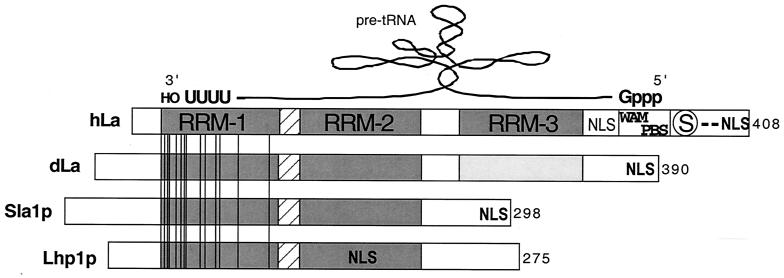
Schematic representation of hLa and its apparent mode of
bipartite interaction with a model precursor tRNA and comparison to
three other La proteins. Cartoon alignment of the La proteins of human
(hLa), D. melanogaster (dLa), S. pombe (Sla1p),
and S. cerevisiae (Lhp1p), representatives of four
phylogenetic branches according to the work of Rosenblum et al.
(117). Note that these represent four polypeptides that
have been characterized as UUU-OH-binding proteins that associate with
nascent Pol III transcripts in vivo (83, 136, 139, 153).
Their lengths, in amino acids, are indicated at the right. RRM-1, -2,
and -3 domains are shaded and labeled for hLa only. Invariant residues
in the 13 sequences are shown as vertical lines; note that these are
limited to RRM-1. The invariant residues (numbered according to hLa)
are Q20, E22, Y24, F25, N29, D33, F35, L36, G45, V47, F55, R57, A71,
and R91. The linker between RRM-1 and RRM-2 is hatched. A putative WAM
and PBS and serine 366 (encircled S) are shown (see text). Positions of
the NLS are indicated in boldface according to reference
. A consensus NLS in hLa, as noted previously
(131), is shown in lightface. A schematized pre-tRNA has
been imposed above the hLa cartoon to illustrate its proposed
orientation according to a model based on prior work (41,
67) as described in the text.

A model of phosphorylation-regulated, bipartite
precursor tRNA binding by the hLa protein. The model shown here depicts
bipartite interactions with a nascent pre-tRNA according to previous
data (41, 67) and the model in Fig. 2. The tandem RRM-1
and -2 of the NTD mediate high-affinity binding to UUU-OH-containing
RNAs. The CTD of hLa contains a basic region and an acidic region,
shown as +++ and −−, respectively; residues 328 to 344 conform to a
WAM, and residues 348 to 368 represent a putative PBS (see Fig. 2 and
text). Together, the WAM and PBS can recognize the 5′-pppG/A motif that
comprises the 5′ ends of nascent Pol III transcripts (41,
67). Serine 366, which resides in a region that demarcates a
transition from basic to acidic residues, is shown as S in
unphosphorylated La and as P in the phosphoserine form.
References
-
- Allain F H, Gilbert D E, Bouvet P, Feigon J. Solution structure of the two N-terminal RNA-binding domains of nucleolin and NMR study of the interaction with its RNA target. J Mol Biol. 2000;303:227–241. - PubMed
-
- Altman S, Kirsebom L. Ribonuclease P. In: Gesteland R F, Chech T R, Atkins J F, editors. The RNA world. 2nd ed. Cold Spring Harbor, N.Y: Cold Spring Harbor Laboratory Press; 1999. pp. 351–380.
Publication types
MeSH terms
Substances
LinkOut - more resources
Full Text Sources
Molecular Biology Databases