. 2001 Jan;21(2):414-24.
doi: 10.1128/MCB.21.2.414-424.2001.

Interleukin-6-induced STAT3 and AP-1 amplify hepatocyte nuclear factor 1-mediated transactivation of hepatic genes, an adaptive response to liver injury

Affiliations

Interleukin-6-induced STAT3 and AP-1 amplify hepatocyte nuclear factor 1-mediated transactivation of hepatic genes, an adaptive response to liver injury

J I Leu et al. Mol Cell Biol. 2001 Jan.

Abstract

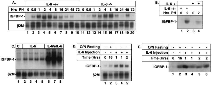
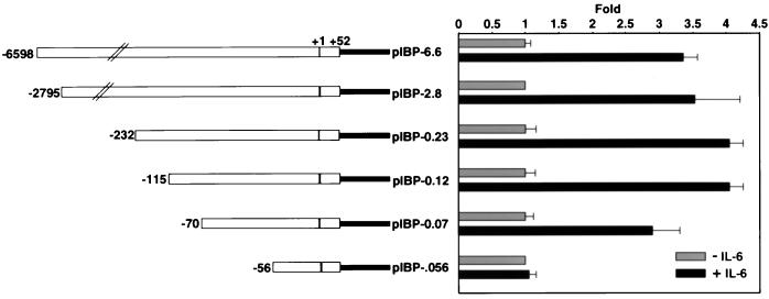
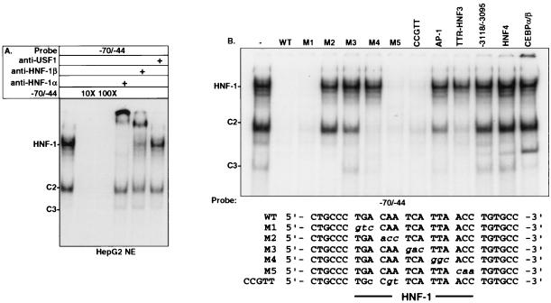
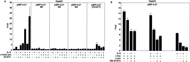
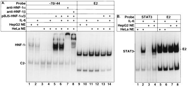
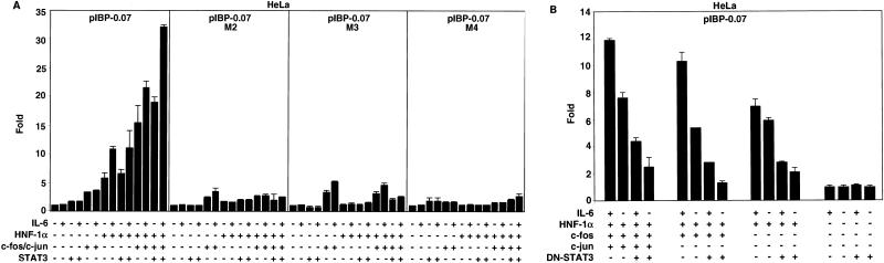
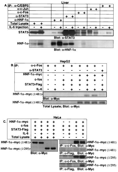
Following hepatic injury or stress, gluconeogenic and acute-phase response genes are rapidly upregulated to restore metabolic homeostasis and limit tissue damage. Regulation of the liver-restricted insulin-like growth factor binding protein 1 (IGFBP-1) gene is dramatically altered by changes in the metabolic state and hepatectomy, and thus it provided an appropriate reporter to assess the transcriptional milieu in the liver during repair and regeneration. The cytokine interleukin-6 (IL-6) is required for liver regeneration and repair, and it transcriptionally upregulates a vast array of genes during liver growth by unknown mechanisms. Evidence for a biologic role of IL-6 in IGFBP-1 upregulation was demonstrated by increased expression of hepatic IGFBP-1 in IL-6 transgenic and following injection of IL-6 into nonfasting animals and its reduced expression in IL-6(-/-) livers posthepatectomy. In both hepatic and nonhepatic cells, IL-6 -mediated IGFBP-1 promoter activation was via an intact hepatocyte nuclear factor 1 (HNF-1) site and was dependent on the presence of endogenous liver factor HNF-1 and induced factors STAT3 and AP-1 (c-Fos/c-Jun). IL-6 acted through the STAT3 pathway, as dominant negative STAT3 completely blocked IL-6-mediated stimulation of the IGFBP-1 promoter via the HNF-1 site. HNF-1/c-Fos and HNF-1/STAT3 protein complexes were detected in mouse livers and in hepatic and nonhepatic cell lines overexpressing STAT3/c-Fos/HNF-1. Similar regulation was demonstrated using glucose-6-phosphatase and alpha-fibrinogen promoters, indicating that HNF-1/IL-6/STAT3/AP-1-mediated transactivation of hepatic gene expression is a general phenomenon after liver injury. These results demonstrate that the two classes of transcription factors, growth induced (STAT3 and AP-1) and tissue specific (HNF-1), can interact as an adaptive response to liver injury to amplify expression of hepatic genes important for the homeostatic response during organ repair.


Figures

FIG. 1
FIG. 1
Enhanced expression of IGFBP-1 in IL-6 transgenic mice, in IL-6+/+ livers posthepatectomy, and after IL-6 treatment. (A) Delayed expression of IGFBP-1 mRNA in IL6−/− livers posthepatectomy (PH). RNA was prepared from IL-6−/− and IL-6+/+ livers at the indicated times after hepatectomy. RNA (10 μg) was gel electrophoresed and probed with nick-translated rat IGFBP-1 cDNA probe. β2-Microglobulin (β2M) was used as a normalizing control. This Northern blot is representative of three. (B) Diminished serum IGFBP-1 protein level in IL-6−/− livers posthepatectomy. Five-microliter aliquots of serum were fractionated on an SDS–12% polyacrylamide gel, blotted, and incubated with the IGFBP-1 antibody. (C) Elevated expression of IGFBP-1 mRNA in IL-6 transgenic and IL-6/IL-6 soluble receptor (IL-6/sIL-6) double-transgenic mice. C, control animal. (D) Induction of hepatic IGFBP-1 mRNA after 16-h (overnight [O/N]) fast or 1 to 2 h after IL-6 injection. The animals were injected subcutaneously with rhIL-6 (1 mg/kg) for 1 or 2 h. (E) Induction of serum IGFBP-1 after overnight fasting or 1 to 2 h after IL-6 treatment.
FIG. 2
FIG. 2
Functional analyses of the mouse IGFBP-1 promoter in HepG2 cells and deletion mapping of the mouse IGFBP-1 promoter. Left, schematic diagrams of the various mouse IGFBP-1 deletion constructs; right, graphical representation of relative luciferase activity after normalization to β-galactosidase activity. To determine enzyme activity, 0.5 μg of the indicated reporter and 1 μg of pRSV-β-galactosidase were transfected in HepG2 cells by the calcium phosphate precipitation method using the 60-mm-diameter dishes. The cells were treated with rhIL-6 (100 ng/ml) for 4 h. The luciferase activity was expressed as fold induction relative to the basal activity of the reporter construct in the absence of IL-6 treatment. Six independent determinants were made for each construct by performing duplicates in three separate experiments. The values were plotted as averages ± standard deviations.
FIG. 3
FIG. 3
Characterization of proteins binding to region −70/−44 of the mouse IGFBP-1 promoter. (A) EMSA using HepG2 nuclear extracts (NE) showing competition and supershift by HNF-1α and -β antibodies. The indicated radiolabeled probe was incubated with HepG2 nuclear extracts in the absence or presence of anti-HNF-1α, anti-HNF-1β, or anti-USF1. For competition assays, a 10- or 100-fold molar excess of unlabeled oligonucleotide of the same sequence was used. Reaction products were fractionated on a 5% nondenaturing acrylamide gel. (B) Effects of selective mutations to HNF-1 site analyzed by EMSA using 100-fold molar excesses of the indicated competitors. Sequences for the wild-type and mutant oligonucleotides between −70 and −44 are shown; sequences for the other competitors are described in Materials and Methods.
FIG. 4
FIG. 4
Regulation of the mouse IGFBP-1 promoter activity by STAT3, c-Fos/c-Jun, and IL-6. (A) An intact HNF-1 binding site coupled with STAT3/c-Fos/c-Jun overexpression in the presence of IL-6 is required for maximal stimulation of the IGFBP-1 promoter in HepG2 cells. (B) Suppression of IL-6-mediated stimulation of the IGFBP-1 promoter via the HNF-1 site by DN-STAT3. HepG2 cells were transfected with the indicated wild-type and mutant pIBP-0.07 constructs (36 ng) in a 24-well plate. For panel A, 36 ng of pCMV-STAT3 with or without 36 ng of pCMV-c-jun and pCMV-c-fos was used. Luciferase activity was expressed as fold induction relative to the basal activity of the reporter construct in the absence of IL-6 treatment and in the absence of the STAT3 or c-Fos/c-Jun expression plasmid. For panel B, 36 ng of pCMV-c-fos with or without 36 ng of pCMV-c-jun or 71 ng of DN-STAT3, as indicated, was used. Luciferase activity was expressed as fold induction relative to the activity of the reporter construct in the presence of DN-STAT3 but in the absence of IL-6 treatment. As indicated, the transfected cells were treated with rhIL-6 (100 ng/ml) for 4 h. Nine independent determinants were made for each construct by performing triplicates in three separate experiments. The values were plotted as averages ± standard deviations.
FIG. 5
FIG. 5
HeLa cells respond to IL-6 and do not contain HNF-1. (A) EMSA using HepG2 and HeLa (NE) with and without 20 min of IL-6 treatment (100 ng/ml) as well as HeLa nuclear extract containing overexpressed HNF-1α and -β in the presence and absence of IL-6 treatment. Anti-HNF-1α and anti-HNF-1β were used in the supershift analyses to verify the overexpressed HNF-1α and -β. (B) HeLa cells contain an intact IL-6 pathway. The indicated radiolabeled probes were incubated with HepG2 and HeLa nuclear extracts (10 μg). Reaction products were fractionated using a 5% nondenaturing acrylamide gel. The E2 probe was used as a normalizing control for loading.
FIG. 6
FIG. 6
Activation of the IGFBP-1 promoter in HeLa cells with HNF-1α and IL-6/STAT3/AP-1 cotransfection. (A) An intact HNF-1 binding site coupled with HNF-1α/STAT3/c-Fos/c-Jun overexpression in the presence of IL-6 is required for maximal stimulation of the IGFBP-1 promoter in HeLa cells. (B) The ability of the exogenous HNF-1 with or without c-Fos or c-Fos/c-Jun to activate the IGFBP-1 promoter in HeLa cells is blocked by DN-STAT3. HeLa cells were transfected with the indicated pIBP-0.07 constructs (29 ng) in 24-well plates. For cotransfection experiments, 29 ng of pCMV-STAT3, 29 ng of pCMV-c-jun, 29 ng of pCMV-c-fos, 29 ng of pcDNA3.1-HNF-1α (aa 1 to 481), or 71 ng of DN-STAT3, as indicated, was used. The transfected cells were treated with rhIL-6 (100 ng/ml) for 4 h. Fold induction relative to the basal activity of the reporter construct in the absence of IL-6 treatment and in the absence of any indicated expression plasmids; fold induction relative to the activity of the reporter construct in the presence of DN-STAT3 but in the absence of IL-6 treatment. Nine independent determinants were made for each construct by performing triplicates in three separate experiments. The values were plotted as averages ± standard deviations.
FIG. 7
FIG. 7
Presence of STAT3/HNF-1α/c-Fos complex in livers, HeLa cells, and HepG2 cells. (A) Coimmunoprecipitation analyses using C57BL/6 mice. For the quiescent time point, the mice were sacrificed by cervical dislocation. For IL-6-treated mice, the animals were injected subcutaneously with rhIL-6 for 30 min. Whole-cell lysates were immunoprecipitated (IP) with the indicated antibodies and analyzed by immunoblotting using STAT3 antibody (top) and HNF-1α antibody (bottom). HepG2 and HeLa cells were transiently transfected with 2 μg of the indicated HNF-1α, 2 μg of pRcCMV-STAT3-Flag, and 2 μg of pCMV-c-fos expression plasmid in 100-mm-diameter plates using the calcium phosphate technique. The cells were kept in 0.2% FBS for 18 h and then incubated with rhIL-6 (100 ng/ml) for 30 min. Whole-cell lysates were immunoprecipitated with the indicated antibodies and analyzed by immunoblotting using the Myc antibody.

References

    1. Babajko S, Groyer A. Interplay of liver-enriched trans-acting factors, DBP and HNF1 in the transactivation of human IGFBP-1 promoter. Biochem Biophys Res Commun. 1993;196:480–486. - PubMed
    1. Babajko S, Tronche F, Groyer A. Liver-specific expression of human insulin-like growth factor binding protein-1: functional role of transcription factor HNF-1 in vivo. Proc Natl Acad Sci USA. 1993;90:272–276. - PMC - PubMed
    1. Baumhueter S, Courtois G, Crabtree G R. A variant nuclear protein in dedifferentiated hepatoma cells binds to the same functional sequences in the β fibrinogen gene promoter as HNF-1. EMBO J. 1988;7:2485–2493. - PMC - PubMed
    1. Cereghini S. Liver-enriched transcription factors and hepatocyte differentiation. FASEB J. 1996;10:267–282. - PubMed
    1. Costa R H, Grayson D R, Xanthopoulos K G, Darnell J E., Jr A liver specific DNA binding protein recognizes multiple nucleotide sites in regulatory regions of transthyretin, α1-antitrypsin, albumin, and simian virus 40 genes. Proc Natl Acad Sci USA. 1988;85:3840–3844. - PMC - PubMed

Publication types

MeSH terms

Substances