. 2001 Jan;21(2):425-37.
doi: 10.1128/MCB.21.2.425-437.2001.

Oligomerization of DH domain is essential for Dbl-induced transformation

Affiliations

Oligomerization of DH domain is essential for Dbl-induced transformation

K Zhu et al. Mol Cell Biol. 2001 Jan.

Abstract

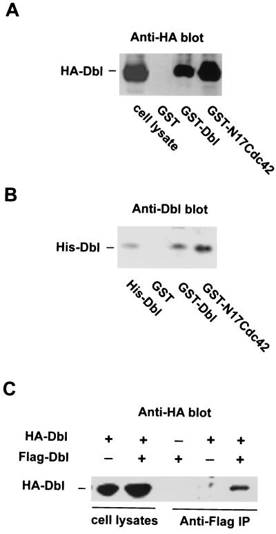
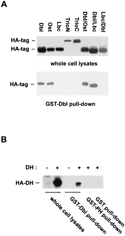
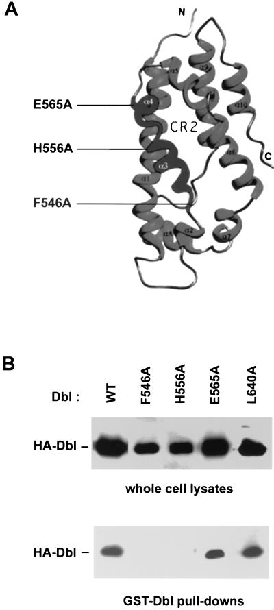
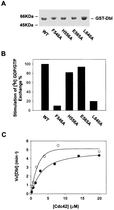
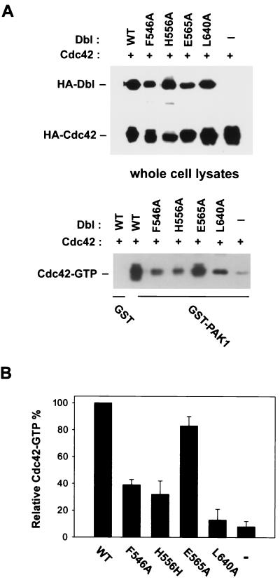
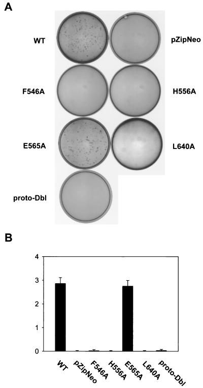
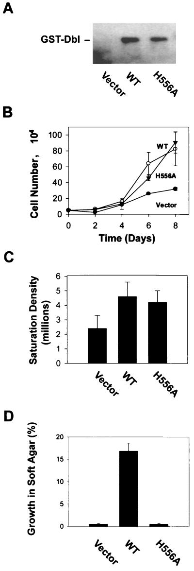
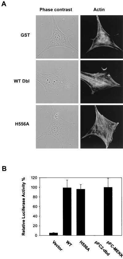
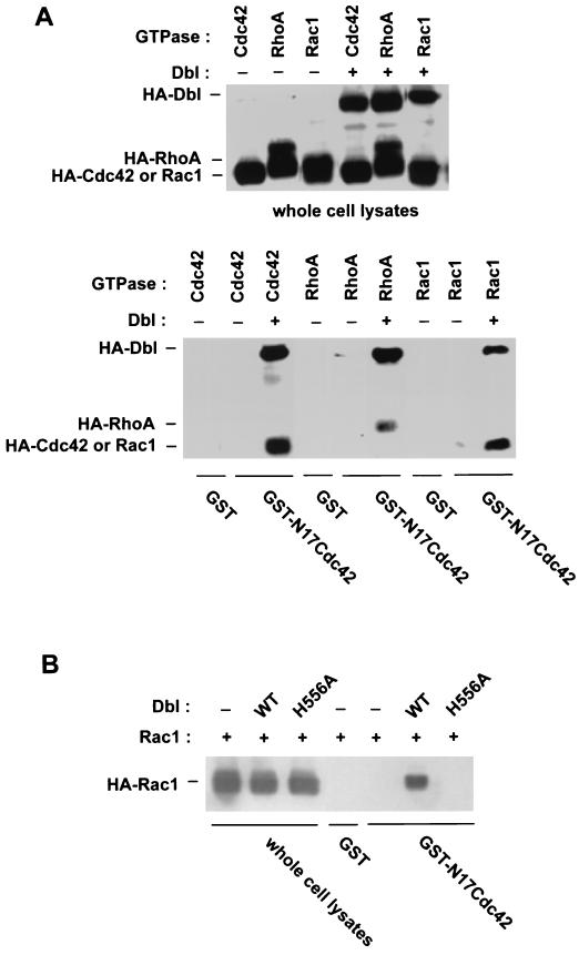
The dbl oncogene product (onco-Dbl) is the prototype member of a family of guanine nucleotide exchange factors (GEFs) for Rho GTPases. The Dbl homology (DH) domain of onco-Dbl is responsible for the GEF catalytic activity, and the DH domain, together with the immediately adjacent pleckstrin homology (PH) domain, constitutes the minimum module bearing transforming function. In the present study, we demonstrate that the onco-Dbl protein exists in oligomeric form in vitro and in cells. The oligomerization is mostly homophilic in nature and is mediated by the DH domain. Mutagenesis studies mapped the region involved in oligomerization to the conserved region 2 of the DH domain, which is located at the opposite side of the Rho GTPase interacting surface. Residue His556 of this region, in particular, is important for this activity, since the H556A mutant retained the GEF catalytic capability and the binding activity toward Cdc42 and RhoA in vitro but was deficient in oligomer formation. Consequently, the Rho GTPase activating potential of the H556A mutant was significantly reduced in cells. The focus-forming and anchorage-independent growth activities of onco-Dbl were completely abolished by the His556-to-Ala mutation, whereas the abilities to stimulate cell growth, activate Jun N-terminal kinase, and cause actin cytoskeletal changes were retained by the mutant. The ability of onco-Dbl to oligomerize allowed multiple Rho GTPases to be recruited to the same signaling complex, and such an ability is defective in the H556A mutant. Taken together, these results suggest that oligomerization of onco-Dbl through the DH domain is essential for cellular transformation by providing the means to generate a signaling complex that further augments and/or coordinates its Rho GTPase activating potential.


Figures

FIG. 1
FIG. 1
Oligomer formation of onco-Dbl. (A) GST-Dbl forms a stable complex with HA-Dbl expressed in Cos-7 cells. HA-Dbl was transiently expressed in Cos-7 cells for 48 h before cells were lysed and analyzed by GST-glutathione affinity precipitation with GST, GST-Dbl, or GST-N17Cdc42. After three washes with ice-cold cell lysis buffer, the coprecipitates were visualized by anti-HA Western blotting. (B) Oligomerization of onco-Dbl is mediated through direct interaction between Dbl molecules. Purified His6-Dbl was incubated with glutathione-agarose-immobilized GST, GST-Dbl, or GST-N17Cdc42 for 30 min before separation by centrifugation. The coprecipitates were detected by Western blotting with anti-Dbl antibody. (C) The oligomerization of onco-Dbl occurs in cells. The Flag-tagged onco-Dbl protein was transiently expressed in Cos-7 cells with or without HA-Dbl, and the cell lysates were subjected to anti-Flag immunoprecipitation followed by anti-HA Western blotting.
FIG. 2
FIG. 2
Oligomerization of onco-Dbl is homophilic in nature and is mediated by the DH domain. (A) Complex formation of GST-Dbl with the HA-tagged Dbl family members and DH-PH chimeras. HA-tagged Dbl, Ost, Lbc, TrioN, TrioC, and the HA-tagged DH domain-PH domain chimeras made between Dbl and Ost (Dbl/Ost), between Dbl and Lbc (Dbl/Lbc), and between Lbc and Dbl (Lbc/Dbl) were expressed in Cos-7 cells. The cell lysates were incubated with glutathione-agarose-immobilized GST-Dbl for 30 min. The GST-Dbl coprecipitates, as well as the input cell lysates, were subjected to anti-HA Western blotting analysis. (B) The DH domain is responsible for complex formation with GST-Dbl. Cell lysates expressing the HA-DH domain of Dbl were subjected to a GST-Dbl, GST-PH, or GST pull-down assay. The input lysates and the glutathione-agarose coprecipitates were visualized by an anti-HA Western blot.
FIG. 3
FIG. 3
Disruption of onco-Dbl oligomer formation by CR2 mutants of the DH domain. (A) Ribbon depiction of the positions of CR2 mutations in the three-dimensional structure of the DH domain. The Rho GTPase interacting CR1 and CR3 are located at the opposite side of CR2. (B) Complex formation of GST-Dbl with the CR2 mutants of onco-Dbl. HA-tagged mutants were transiently expressed in Cos-7 cells. The GST-Dbl coprecipitates from the cell lysates, as well as the input cell lysates, were visualized by an anti-HA Western blot. The L640A mutation is located at the center of CR3. WT, wild type.
FIG. 4
FIG. 4
Effect of CR2 mutations on the GEF activity of onco-Dbl. The cDNAs encoding the wild-type DH-PH module or the module bearing mutations in the DH domain were cloned into an insect cell transfer vector with an N-terminal GST fusion tag for functional expression in Sf9 insect cells. (A) Coomassie blue-stained SDS-PAGE gel of the insect cell-expressed, glutathione-agarose affinity-purified GST fusion mutants. WT, wild type. (B) Relative GEF activities of the recombinant DH domain mutants on RhoA. Approximately 0.2 μg of purified GST-Dbl or Dbl mutant was incubated with 1 μg of [3H]GDP-loaded RhoA in the GEF reaction buffer for 5 min before termination of the reaction by nitrocellulose filtration. The percent retention of RhoA-bound [3H]GDP catalyzed by the mutants was normalized to that catalyzed by wild-type Dbl. (C) Derivation of the kinetic parameters of wild-type Dbl and the H556A mutant using Cdc42 as substrate. The V0s were determined in the presence of 20 nM Dbl or H556A mutant at 1-min intervals with various concentrations of Cdc42-GDP. The resulting V0 and substrate concentration data were best fitted into a modified Michaelis-Menton equation, with corrections being made for basal GDP dissociation from Cdc42.
FIG. 5
FIG. 5
Interaction of the DH mutants with dominant-negative RhoA. Wild-type Dbl and various DH mutants were expressed as HA-tagged proteins in Cos-7 cells by transient transfection. Glutathione-agarose-immobilized GST or GST-N19RhoA (5 μg/sample) was incubated with the respective Cos-7 cell lysates for 1 h followed by centrifugation and three washes. The expression of the respective HA-DH mutants in the cell lysates and their coprecipitation patterns with GST or GST-N19RhoA were detected by anti-HA Western blotting. WT, wild type.
FIG. 6
FIG. 6
Cdc42 activation potential of the DH mutants in cells. (A) The HA-tagged wild-type Dbl or the DH mutants were cotransfected with HA-tagged Cdc42 in Cos-7 cells. At 48 h posttransfection, cell lysates were subjected to GST-PAK1 affinity precipitation. The coprecipitated Cdc42-GTP was detected by anti-HA Western blotting. A sample that was 10% of the amount of whole cell lysates used for GST-PAK1 incubations was also subjected to anti-HA blotting in parallel. WT, wild type. (B) Quantification of the Cdc42-GTP pull-down assays by densitometry measurement. The amount of Cdc42-GTP coprecipitate for the wild-type Dbl cotransfected cells was treated as 100%. The data represent results from four independent experiments.
FIG. 7
FIG. 7
Effect of DH mutations on the transforming activity of Dbl. cDNAs encoding the wild-type DH-PH domain module of Dbl (residues 498 to 825), the DH mutation-bearing DH-PH modules, and proto-Dbl were subcloned into the pZipneoGST vector and assayed for focus-forming activity in NIH 3T3 cells. Foci were quantified at 14 days posttransfection by Giemsa staining. WT, wild type. (A) Tissue culture dishes transfected with 0.1 μg of pZipneoGST-Dbl cDNA were visualized directly by a video camera. (B) Normalized focus-forming activities (103 foci/μg of DNA) of the DH mutants made in CR2 compared to those of wild-type Dbl, proto-Dbl, and the CR3 L640A mutant.
FIG. 8
FIG. 8
Growth properties of the H556A mutant-expressing NIH 3T3 cells. (A) Mock-transfected NIH 3T3 cells (pZipneoGST vector) and the cell clones stably expressing wild-type (WT) GST-Dbl or GST-H556A were analyzed by anti-GST Western blotting. (B) The cell growth rate of the H559A mutant-expressing cells was compared with that of wild-type Dbl-expressing or mock-transfected cells. Cell growth was initiated at a density of 5,000/35-mm-diameter culture dish at day 0 in DMEM supplemented with 2% calf serum. The number of cells in the dishes was counted in 2-day intervals. (C) Cells were plated at a density of 50,000/100-mm-diameter dish at day 0. The saturation densities of the cells were determined after the cell growth was stopped, at day 9. (D) The ability of the transfectants to grow on soft agar was measured in DMEM supplemented with 10% calf serum and 0.3% agarose on top of solidified DMEM with 0.5% agarose. Colonies were scored at 3 weeks postplating under a microscope.
FIG. 9
FIG. 9
Effect of the oligomer-deficient H556A mutant on actin cytoskeletal structure and JNK activation. (A) The morphology and actin structures of H556A mutant-or wild-type (WT) Dbl-expressing NIH 3T3 cells, as well as of the mock-transfected cells, were visualized under a phase-contrast or fluorescence microscope after actin staining with rhodamine-conjugated phalloidin. (B) Various pKH3 constructs (0.4 μg) or controls (pFC-MEKK and pFC2-dbd plasmids) (0.1 μg) were transiently cotransfected into NIH 3T3 cells together with the pFR-Luc reporter plasmid (1 μg) and pFA2-cJun plasmid (0.1 μg). At 48 h posttransfection, the cells were washed and harvested for the measurement of luciferase activities.
FIG. 10
FIG. 10
The oligomeric complex of onco-Dbl is capable of recruiting multiple Rho GTPases. (A) HA-tagged Cdc42, RhoA, or Rac1 was expressed in Cos-7 cells alone or together with HA-Dbl. The cell lysates were incubated with glutathione-agarose-immobilized GST or GST-N17Cdc42 for 30 min. The input cell lysates and the GST fusion coprecipitates were analyzed in parallel by anti-HA Western blotting. (B) HA-Rac1 was expressed alone, together with wild-type (WT) onco-Dbl, or together with the H556A mutant of onco-Dbl in Cos-7 cells. The cell lysates and the GST or GST-N17Cdc42 coprecipitates from the cell lysates were probed with anti-HA antibody in a Western blot.

References

    1. Abe K, Whitehead I P, O'Bryan J P, Der C J. Involvement of NH2-terminal sequences in the negative regulation of Vav signaling and transforming activity. J Biol Chem. 1999;274:30410–30418. - PubMed
    1. Aghazadeh B, Zhu K, Kubiseski T J, Liu G A, Pawson T, Zheng Y, Rosen M K. Structure and mutagenesis of the Dbl homology domain. Nat Struct Biol. 1998;12:1098–1107. - PubMed
    1. Aghazadeh B, Lowry W E, Huang X-Y, Rosen M K. Structural basis for relief of autoinhibition of the Dbl homology domain of proto-oncogene Vav by tyrosine phosphorylation. Cell. 2000;102:625–633. - PubMed
    1. Anborgh P H, Qian X, Papageorge A G, Vass W C, DeClue J E, Lowy D R. Ras-specific exchange factor GRF: oligomerization through its Dbl homology domain and calcium-dependent activation of Raf. Mol Cell Biol. 1999;19:4611–4622. - PMC - PubMed
    1. Boriack-Sjodin P A, Margarit S M, Bar-Sagi D, Kuriyan J. The structural basis of the activation of Ras by Sos. Nature. 1998;394:337–343. - PubMed

Publication types

MeSH terms

Substances