. 2001 Jan 2;98(1):93-8.
doi: 10.1073/pnas.98.1.93.

Constitutive arrestin-mediated desensitization of a human vasopressin receptor mutant associated with nephrogenic diabetes insipidus

Affiliations

Constitutive arrestin-mediated desensitization of a human vasopressin receptor mutant associated with nephrogenic diabetes insipidus

L S Barak et al. Proc Natl Acad Sci U S A. .

Abstract

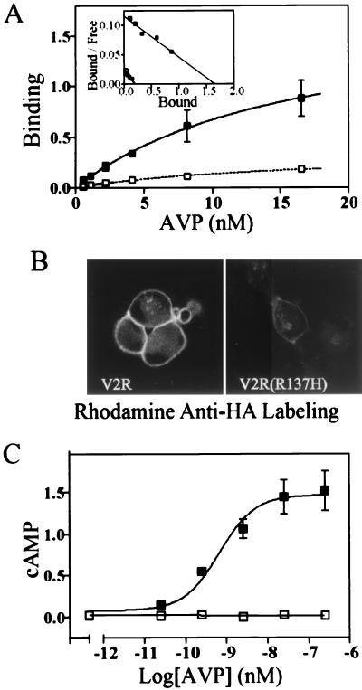
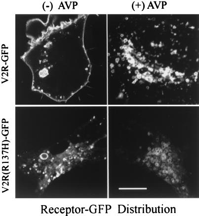
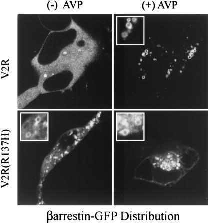
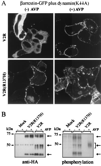
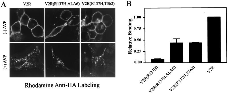
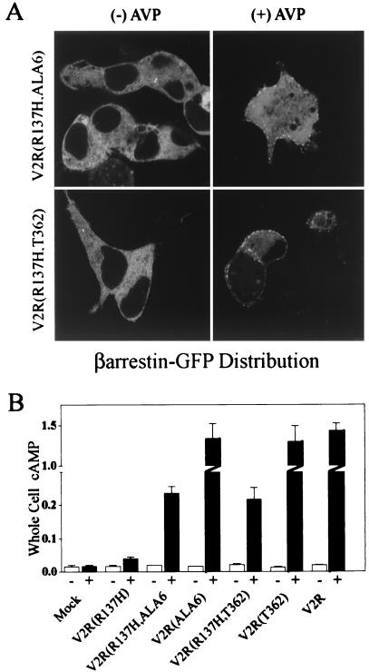
Agonist-dependent desensitization and internalization of G protein-coupled receptors (GPCR) are mediated by the binding of arrestins to phosphorylated receptors. The affinity of arrestins for the phosphorylated GPCR regulates the ability of the internalized receptor to be dephosphorylated and recycled back to the plasma membrane. In this study, we show that the naturally occurring loss of function vasopressin receptor mutation R137H, which is associated with familial nephrogenic diabetes insipidus, induces constitutive arrestin-mediated desensitization. In contrast to the wild-type vasopressin receptor, the nonsignaling R137H receptor is phosphorylated and sequestered in arrestin-associated intracellular vesicles even in the absence of agonist. Eliminating molecular determinants on the receptor that promote high affinity arrestin-receptor interaction reestablishes plasma membrane localization and the ability of the mutated receptors to signal. These findings suggest that unregulated desensitization can contribute to the etiology of a GPCR-based disease, implying that pharmacological targeting of GPCR desensitization may be therapeutically beneficial.


Figures

Figure 1
Figure 1
Expression and adenylyl cyclase stimulation of V2R and V2R(R137H) in HEK-293 cells. Cells transiently transfected with cDNA for V2R (■) or V2R(R137H) (□) were exposed to varying concentrations of [3H]AVP. (A) Scatchard analysis (Inset) indicates the receptors have similar affinity for AVP [V2R, 16 ± 6 nM; V2R(R137H), 15 ± 3 nM]. V2R expression varied between 2.5 and 5 pmol/mg of cell protein, with the plasma membrane expression of the V2R(R137H) being approximately 1/12 of this (x intercept of Scatchard). The data are representative of three experiments, with each point measured in duplicate. (B) Fluorescence images of live unpermeabilized cells, labeled with rhodamine-tagged mouse-monoclonal anti-HA antibodies, expressing the V2R (Left) or the V2R(R137H) (Right). (C). cAMP measured in whole cells stimulated for 15 min with concentrations of AVP between 0 and 250 nM. cAMP accumulation is expressed as total counts of [3H]cAMP/total uptake of [3H]adenine per well of cells. Results are the mean ± SD of three experiments.
Figure 2
Figure 2
Fluorescence images of V2R-GFP and V2R(R137H)-GFP in HEK-293 cells. Cells expressing V2R-GFP (Upper) or V2R(R137H)-GFP (Lower) were treated with vehicle or AVP for 30 min at 37°C. The agonist-mediated redistribution of wild-type receptor (Upper) from the plasma membrane to endocytic vesicles contrasts with the agonist-independent localization of the V2R(R137H)-GFP in endosomes (Lower). Bar = 25 μM.
Figure 3
Figure 3
Fluorescence images of the association between βarrestin2-GFP and V2R or V2R(R137H) in HEK-293 cells. Left shows the agonist-independent distribution of βarrestin2-GFP in living cells expressing the V2R (Upper) or V2R(R137H) (Lower). Without agonist treatment, βarrestin2-GFP is cytosolic in cells containing wild-type V2R (Left Upper), but in cells containing the V2R(R137H) (Left Lower) it is also in endocytic vesicles. Following 30 min of exposure to 100 nM AVP (Right), βarrestin-GFP is localized on endocytic vesicles with both receptor subtypes.
Figure 4
Figure 4
βarrestin2 association with and phosphorylation of V2R and V2R(R137H) in HEK-293 cells. (A) Dynamin(K44A) was expressed with βarrestin2-GFP and either V2R or V2R(R137H). Exposure of V2R to AVP (100 nM) (Upper Right) resulted in appreciable βarrestin2-GFP translocation that remains visible at 30 min as a punctate distribution at the cell membrane rather than as a vesicular distribution inside the cell. In the absence of agonist, the cells containing dynamin(K44A) and V2R(R137H) (Lower Left) also show βarrestin2-GFP fluorescence distributed in punctate areas at the plasma membrane. A similar pattern was apparent even in the presence of agonist (Lower Right). (B) Left shows receptors that were immunoprecipitated with a mouse anti-HA antibody and blotted with a rabbit anti-HA antibody. The faint 50-kDa band present in all six lanes is cross-reactive mouse Ig heavy chain. Right shows depicts receptors that were assayed for phosphorylation as described in Experimental Procedures. Equal amounts of receptor (40 fmol) were loaded into each lane. The arrows mark the positions of the receptor species migrating at approximately 70, 50, and 40 kDa as revealed by anti-HA antibody. Results are representative of three experiments.
Figure 5
Figure 5
Expression of V2R, V2R(R137H,Ala6), and V2R(R137H,T362) in HEK-293 cells. (A) Plasma membrane receptors were labeled with rhodamine-tagged mouse-monoclonal anti-HA antibody. The upper panels show receptor distribution in the absence of agonist. The lower panels show cells that were labeled with antibody before treatment with 100 nM AVP for 30 min at 37°C. (B) Plasma membrane receptor expression measured by [3H]AVP was normalized to wild-type V2R plasma membrane expression (approximately 5 pmol/mg). Data are expressed as the mean ± SD from three independent experiments.
Figure 6
Figure 6
βarrestin2-GFP translocation and adenylyl cyclase response of V2R, V2R(R137H,Ala6), and V2R(R137H,T362) in HEK-293 cells. (A) In the absence of agonist (Left), GFP fluorescence is cytosolic. Following exposure of the cells to 100 nM AVP for 30 min at 37°C, GFP fluorescence redistributes to punctate areas of plasma membrane (Right). (B) Cells were treated with vehicle or 2.5 μM AVP for 15 min, and whole cell cAMP was determined as described in Experimental Procedures. The absolute basal and stimulated cAMP responses were presented in units of (counts of [3H]cAMP per min per well)/(counts of [3H]adenine uptake per minute per well) and were mock (0.019 ± 0.009, 0.017 ± 0.006, n = 3); V2R(R137H) (0.018 ± 0.0045, 0.040 ± 0.008, n = 4); V2R(R137H,Ala6) (0.020 ± 0.0017, 0.23 ± 0.040, n = 4); V2R(Ala6) (0.017 ± 0.0006, 1.3 ± 0.31, n = 3); V2R(R137H,T362) (0.022 ± 0.0056, 0.22 ± 0.070, n = 4); V2R(T362) (0.015 ± 0.0021, 1.3 ± 0.34, n = 3); and V2R (0.021 ± 0.0024, 1.4 ± 0.19, n = 4). Data are expressed as the mean ± SD of three to four separate experiments.

Similar articles

Cited by

References

    1. Ferguson S S, Barak L S, Zhang J, Caron M G. Can J Physiol Pharmacol. 1996;74:1095–1110. - PubMed
    1. Krupnick J G, Benovic J L. Annu Rev Pharmacol Toxicol. 1998;38:289–319. - PubMed
    1. Attramadal H, Arriza J L, Aoki C, Dawson T M, Codina J, Kwatra M M, Snyder S H, Caron M G, Lefkowitz R J. J Biol Chem. 1992;267:17882–17890. - PubMed
    1. Gurevich V V, Dion S B, Onorato J J, Ptasienski J, Kim C M, Sterne-Marr R, Hosey M M, Benovic J L. J Biol Chem. 1995;270:720–731. - PubMed
    1. Barak L S, Ferguson S S, Zhang J, Caron M G. J Biol Chem. 1997;272:27497–27500. - PubMed

Publication types

MeSH terms