. 2001 Jan 2;98(1):301-6.
doi: 10.1073/pnas.98.1.301.

Dopamine D1/D5 receptor modulation of excitatory synaptic inputs to layer V prefrontal cortex neurons

Affiliations

Dopamine D1/D5 receptor modulation of excitatory synaptic inputs to layer V prefrontal cortex neurons

J K Seamans et al. Proc Natl Acad Sci U S A. .

Abstract

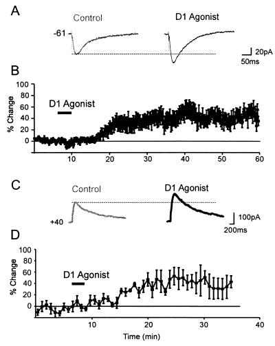
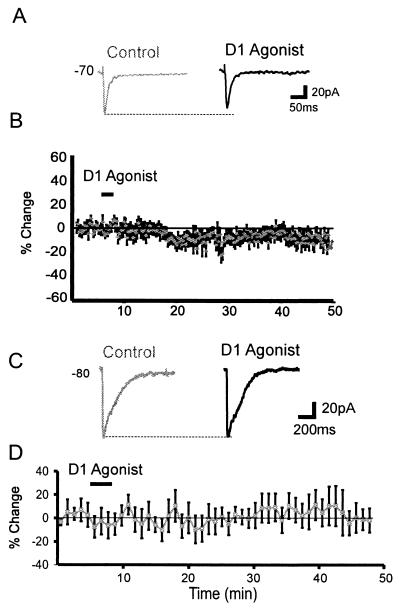
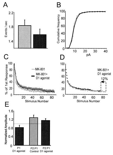
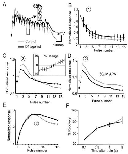
Dopamine acts mainly through the D1/D5 receptor in the prefrontal cortex (PFC) to modulate neural activity and behaviors associated with working memory. To understand the mechanism of this effect, we examined the modulation of excitatory synaptic inputs onto layer V PFC pyramidal neurons by D1/D5 receptor stimulation. D1/D5 agonists increased the size of N-methyl-d-aspartate (NMDA) component of excitatory postsynaptic currents (EPSCs) through a postsynaptic mechanism. In contrast, D1/D5 agonists caused a slight reduction in the size of the non-NMDA component of EPSCs through a small decrease in release probability. With 20 Hz synaptic trains, we found that the D1/D5 agonists increased depolarization of summating the NMDA component of excitatory postsynaptic potential (EPSP). By increasing the NMDA component of EPSCs, yet slightly reducing release, D1/D5 receptor activation selectively enhanced sustained synaptic inputs and equalized the sizes of EPSPs in a 20-Hz train.


Figures

Figure 1
Figure 1
D1 receptor modulation of NMDA responses in PFC neurons. (A) Representative synaptic responses. For all traces, the baseline (control) response is shown at left, the response during the period of the peak D1 agonist response (>10 min after D1 agonist offset) is on the right. A D1 agonist (0.5 μM SKF81297) enhanced the NMDA EPSC. (B) Average percentage change (and SEM) in NMDA EPSC amplitude over time. (C) Representative traces showing that the response evoked by puffing NMDA (1 mM, 8 ms) was enhanced by the D1 agonist (10 μM SKF-81297, black trace) relative to the baseline response (gray traces) at Vhold = +40 mV. (D) Average percentage change (and SEM) in postsynaptic NMDA current amplitude (n = 8).
Figure 2
Figure 2
D1 receptor modulation of non-NMDA responses in PFC neurons. (A) Representative traces showing that a D1 agonist (1 μM SKF81297) slightly reduced the non-NMDA EPSC. (B) Average percentage change (and SEM) in non-NMDA EPSC amplitude over time. (C) Representative traces showing the non-NMDA response evoked by puffing glutamate (1 mM, 8 ms) in the presence of APV (100 μM) was unaffected by the D1 agonist (10 μM SKF-81297, black trace) relative to the baseline response (gray traces) at Vhold = −80 mV. TTX (500 nM) was also included in the bathing solution and CsCl containing pipettes were used. (D) Average percentage change (and SEM) in postsynaptic non-NMDA current amplitude (n = 8).
Figure 3
Figure 3
Presynaptic modulation by D1 agonists. (A) Histogram of group data showing the mean mEPSC frequency for a 300-s period for the control (gray) and D1 agonist condition (black). The drop in mEPSC frequency in the D1 agonist condition was most evident for the first two bins. (B) The same data as in A replotted as a cumulative histogram. For A and B, mEPSCs were recorded at −70 mV in 0.25–1 μM TTX and 10–20 μM bicuculline by using CsCl-filled pipettes. Data were analyzed for the 5 min before D1 agonist application and 10–15 min after D1 agonist offset. (C) The MK-801 blocking function for control cells (gray boxes, n = 7) and for cells receiving the D1 agonist (▴, n = 6). (D) The D1 agonist blocking function overlaps with that of the control if the D1 agonist blocking function was accelerated by 12% by shifting the x axis. (E) Histogram representing data from paired pulse experiments. (Left, black bar) The normalized change in response 1 (P1) in the 10–40 min following application of the D1 agonist. (Middle, gray bar) The ratio of P2:P1 for the control condition. (Right, black bar) The ratio of P2:P1 for responses acquired 10–40 min following application of the D1 agonist. Pulses were separated by 20 ms, and neurons were recorded under voltage–clamp in the presence of 10 μM bicuculline and 0.5–2 μM DNQX by using CsCl/QX-314-filled pipettes.
Figure 4
Figure 4
Modulation of synaptic depression by D1 agonists. (A) Representative trace showing the nonisolated synaptic response to 20-Hz trains under control conditions (gray traces) and following a D1 agonist (10 μM SKF-81297, black traces). Responses were obtained by using QX-314-filled patch-pipettes in 0.5 μM bicuculline. The inset shows the procedure for measuring response size. Measurement 1 assessed the size of each individual EPSP from a baseline obtained just before each pulse in the train, whereas measurement 2 assessed the size of each response plus the concomitant depolarization on which the EPSP was riding by calculating the integral under each EPSP relative to the initial baseline. (B) Average normalized amplitude (and SEM) by using measurement 1 for the control period (gray line) and following a D1 agonist (black line). The lines represent the fit to the data by using a single exponential function. (C) Average normalized amplitude by using measurement 2 for the control period (gray line) and following a D1 agonist (black line). The D1 agonist condition was normalized to the initial averaged response for the control condition. (Inset) The same data replotted as percent change relative to the control response (n = 10). (D) In an additional group of six cells recorded in the presence of APV, no such cross over was observed following application of a D1 agonist. (C and D) The lines connecting points 1 and 2 in each figure are dotted to separate the initial facilitation from depression. (E) When NMDA receptors carried the majority of the synaptic current by conducting experiments in CNQX (3 μM) and bicuculline (10 μM). D1 agonists also caused a relative increase in later responses in the train. (F) Recovery assessed by single pulses delivered 0.1, 0.5,1, 5s after the 15-pulse/20-Hz train was unaffected by D1 agonists.

References

    1. Watanabe M, Kodama T, Hikosaka K. J Neurophysiol. 1997;78:2795–2798. - PubMed
    1. Sawaguchi T, Matsumura M, Kubota K. J Neurophysiol. 1990;63:1385–1400. - PubMed
    1. Sawaguchi T, Matsumura M, Kubota K. J Neurophysiol. 1990;63:1401–1412. - PubMed
    1. Williams G V, Goldman-Rakic P S. Nature (London) 1995;376:572–575. - PubMed
    1. Seamans J K, Floresco S B, Phillips A G. J Neurosci. 1998;18:1613–1621. - PMC - PubMed

Publication types

MeSH terms

LinkOut - more resources