. 2001 Jan 2;98(1):194-9.
doi: 10.1073/pnas.98.1.194.

Deletion of the p27Kip1 gene restores normal development in cyclin D1-deficient mice

Affiliations

Deletion of the p27Kip1 gene restores normal development in cyclin D1-deficient mice

Y Geng et al. Proc Natl Acad Sci U S A. .

Abstract

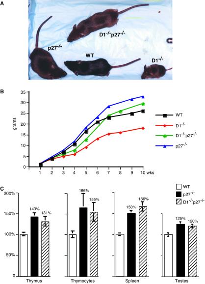
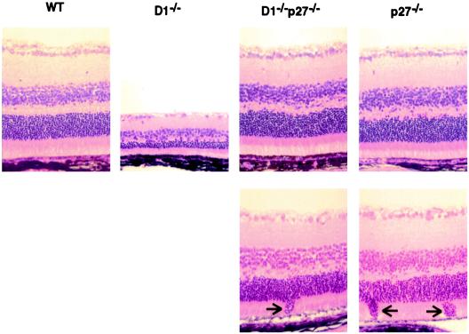
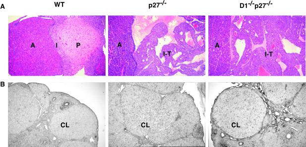
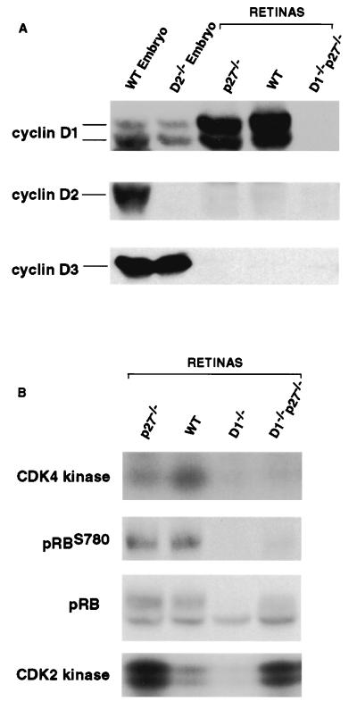
D-type cyclins (cyclins D1, D2, and D3) are key components of cell cycle machinery in mammalian cells. These proteins are believed to drive cell cycle progression by associating with their kinase partners, cyclin-dependent kinases, and by directing phosphorylation of critical cellular substrates. In addition, D-cyclins play a kinase-independent role by sequestering cell cycle inhibitors p27(Kip1) and p21(Cip1). In the past, we and others generated cyclin D1-deficient mice and have shown that these mice display developmental abnormalities, hypoplastic retinas, and pregnancy-insensitive mammary glands. To test the significance of cyclin D1-p27(Kip1) interaction within a living mouse, we crossed cyclin D1-deficient mice with mice lacking p27(Kip1), and we generated double-mutant cyclin D1(-/-)p27(-/-) animals. Here we report that ablation of p27(Kip1) restores essentially normal development in cyclin D1-deficient mice. Our results provide genetic evidence that p27(Kip1) functions downstream of cyclin D1.


Figures

Figure 1
Figure 1
Impact of the loss of cyclin D1 and p27Kip1 on animal growth. (A) A photograph of 8-week-old wild-type (WT) and mutant males. (B) Growth curves of wild-type (WT) and mutant males. The mean body weight values were obtained by weekly weighing 6–15 animals per experimental group. An identical analysis was performed for females, and very similar curves were obtained (not shown). (C) Relative organ weight and the number of cells per thymus in wild type (WT) and mutant males. For each genotype, five 10- to 13-week-old males were analyzed. The weight of indicated organs or the total number of cells per thymus (thymocytes) was determined and divided by animal body weight. The mean values shown are expressed relative to the values seen in wild-type mice, which were set at 100%. Error bars indicate SE.
Figure 2
Figure 2
Ablation of p27Kip1 rescues the retinal phenotype of cyclin D1-deficient mice. Histologic sections of retinas derived from wild-type (WT) or mutant mice were stained with hematoxylin and eosin. (Lower) Focal invasions of the outer granular layer into the layer of rods and cones, seen in p27−/− and cyclin D1−/−p27−/− mice, are indicated by arrows.
Figure 3
Figure 3
Ablation of p27Kip1 rescues the mammary epithelial phenotype of cyclin D1-deficient mice. A wild-type (WT) or mutant mammary epithelial tree 1 day after the delivery of pups. Mammary whole mounts were stained with carmine red. Mammary epithelial transplantations were performed as described in Materials and Methods.
Figure 4
Figure 4
Ablation of cyclin D1 does not rescue the phenotype of p27Kip1-deficient mice. (A) Histologic sections of pituitaries derived from 10- to 13-week-old wild-type (WT) or mutant mice were stained with hematoxylin and eosin. Note the normal appearance of pituitaries in WT mice and the presence of intermediate lobe tumors (I-T) in p27−/− and cyclin D1−/−p27−/− animals. A, anterior lobe of the pituitary; P, posterior lobe; I, intermediate lobe. (B) Incorporation of BrdUrd into corpora lutea (CL) of wild-type (WT) and mutant ovaries 3 days after superovulation. Mutant p27−/− and cyclin D1−/−p27−/− corpora lutea continue to incorporate BrdUrd, in contrast to wild-type ovaries.
Figure 5
Figure 5
Molecular analyses of mutant retinas. Retinal lysates were prepared from mice of indicated genotypes. The levels of D-cyclins and serine 780 phospho-Rb (pRBS780) and total levels of pRB were determined by Western blotting. CDK4-associated or CDK2-associated kinase activity was determined by immunoprecipitation–kinase assays.

References

    1. Sherr C J. Trends Biochem Sci. 1995;20:187–190. - PubMed
    1. Bartkova J, Lukas J, Strauss M, Bartek J. Oncogene. 1995;10:775–778. - PubMed
    1. Motokura T, Bloom T, Kim H G, Juppner H, Ruderman J V, Kronenberg H M, Arnold A. Nature (London) 1991;350:512–515. - PubMed
    1. Dickson C, Fantl V, Gillett C, Brookes S, Bartek J, Smith R, Fisher C, Barnes D, Peters G. Cancer Lett. 1995;90:43–50. - PubMed
    1. Weinstat-Saslow D, Merino M J, Manrow R E, Lawrence J A, Bluth R F, Wittenbel K D, Simpson J F, Page D L, Steeg P S. Nat Med. 1995;1:1257–1260. - PubMed

Publication types

MeSH terms

LinkOut - more resources