. 2001 Jan 2;98(1):136-41.
doi: 10.1073/pnas.98.1.136.

A role of the kinase mTOR in cellular transformation induced by the oncoproteins P3k and Akt

Affiliations

A role of the kinase mTOR in cellular transformation induced by the oncoproteins P3k and Akt

M Aoki et al. Proc Natl Acad Sci U S A. .

Abstract

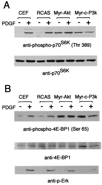
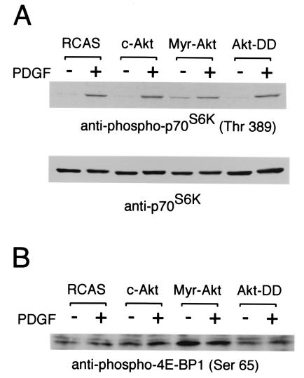
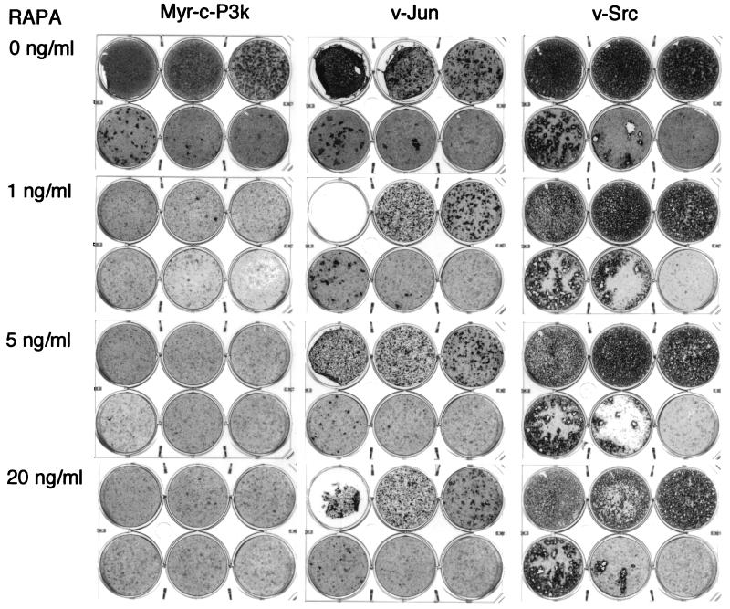
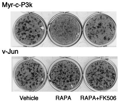
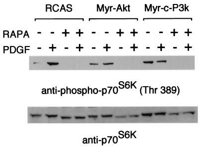
The oncoproteins P3k (homolog of the catalytic subunit of class IA phosphoinositide 3-kinase) and Akt (protein kinase B) induce oncogenic transformation of chicken embryo fibroblasts. The transformed cells show constitutive phosphorylation of the positive regulator of translation p70S6 kinase (S6K) and of the eukaryotic initiation factor 4E-BP1 binding protein (4E-BP1), a negative regulator of translation. Phosphorylation activates S6K and inactivates 4E-BP1. A mutant of Akt that retains kinase activity but does not induce phosphorylation of S6K or of 4E-BP1 fails to transform chicken embryo fibroblasts, suggesting a correlation between the oncogenicity of Akt and phosphorylation of S6K and 4E-BP1. The macrolide antibiotic rapamycin effectively blocks oncogenic transformation induced by either P3k or Akt but does not reduce the transforming activity of 11 other oncoproteins. Rapamycin inhibits the kinase mTOR, an important regulator of translation, and this inhibition requires binding of the antibiotic to the immunophilin FKBP12. Displacement of rapamycin from FKBP12 relieves the inhibition of mTOR and also restores P3k-induced transformation. These data are in accord with the hypothesis that transformation by P3k or Akt involves intervention in translational controls.


Figures

Figure 1
Figure 1
Constitutive phosphorylation of S6K and 4E-BP1 in CEF transformed with P3k and Akt. CEF and CEF infected with RCAS, RCAS-Myr-Akt, and RCAS-Myr-c-P3k were serum starved for 40 h and then stimulated with PDGF for 15 min. The cells were lysed, and the lysates were resolved in a 10% (A) or a 15% (B) SDS-polyacrylamide gel and then transferred to a polyvinylidene difluoride membrane. The blot was probed with anti-phospho-S6K (threonine 389), anti-S6K, anti-phospho-4E-BP1 (serine 65), anti-4E-BP1, or anti-phospho-Erk antibody.
Figure 2
Figure 2
Correlation between the ability to induce S6k activation and 4E-BP1 inactivation with the ability to induce oncogenic transformation. Cell lysates were prepared as described in the legend to Fig. 1. The lysates were resolved in a 10% (A) or a 15% (B) SDS-polyacrylamide gel. The blot was probed with anti-phospho-S6K (threonine 389), anti-S6K, or anti-phospho-4E-BP1 (serine 65) antibody.
Figure 3
Figure 3
Rapamycin inhibits focus formation by P3k but not Jun or Src. CEF were infected with viruses containing the indicated oncoproteins. Each plate was infected with 100 μl of the virus stocks diluted to 10−1 (top left well), 10−2 (top center well), 10−3 (top right well), 10−4 (bottom left well), 10−5 (bottom center well), or with no viruses (bottom right well). The cells were overlaid with nutrient agar containing the indicated concentrations of rapamycin (RAPA) for 17 days and then fixed and stained with crystal violet.
Figure 4
Figure 4
Rapamycin inhibition of the P3k-induced transformation of CEF is FKBP 12 dependent. CEF were infected with viruses and then overlaid with nutrient agar containing 1 ng/ml rapamycin (Center), 1 ng/ml rapamycin and 100 molar excess of FK506 (Right), or vehicle (DMSO, Left) only.
Figure 5
Figure 5
Rapamycin inhibits phosphorylation of S6K. CEF were serum starved for 40 h and treated or not treated with rapamycin (RAPA, 10 ng/ml) for 2 h and then stimulated with PDGF (50 ng/ml) for 15 min. The cell lysates were prepared and resolved in a 10% SDS-polyacrylamide gel. The blot was probed with anti-phospho-S6K or with anti-S6K antibody.

References

    1. Bellacosa A, Testa J R, Staal S P, Tsichlis P N. Science. 1991;254:274–277. - PubMed
    1. Chang H W, Aoki M, Fruman D, Auger K R, Bellacosa A, Tsichlis P N, Cantley L C, Roberts T M, Vogt P K. Science. 1997;276:1848–1850. - PubMed
    1. Shepherd P R, Withers D J, Siddle K. Biochem J. 1998;333:471–490. - PMC - PubMed
    1. Alessi D R, Downes C P. Biochim Biophys Acta. 1998;1436:151–164. - PubMed
    1. Fruman D A, Meyers R E, Cantley L C. Annu Rev Biochem. 1998;67:481–507. - PubMed

Publication types

MeSH terms

LinkOut - more resources