. 2001 Jan 16;98(2):415-20.
doi: 10.1073/pnas.98.2.415. Epub 2000 Dec 26.

Coupling of histone deacetylation to NAD breakdown by the yeast silencing protein Sir2: Evidence for acetyl transfer from substrate to an NAD breakdown product

Affiliations

Coupling of histone deacetylation to NAD breakdown by the yeast silencing protein Sir2: Evidence for acetyl transfer from substrate to an NAD breakdown product

J C Tanny et al. Proc Natl Acad Sci U S A. .

Abstract

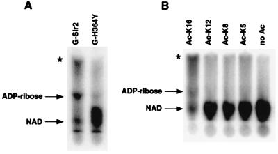
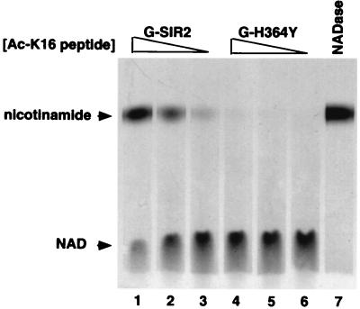
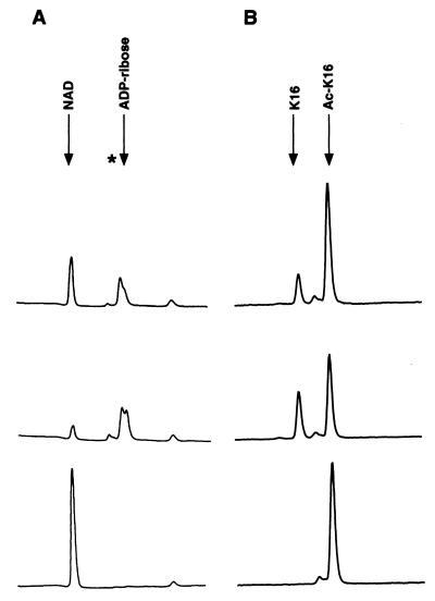
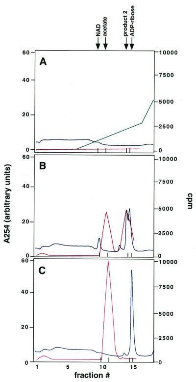
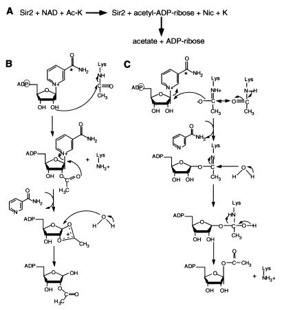
The Saccharomyces cerevisiae silencing protein Sir2 is the founding member of a universally conserved family of proteins that have been shown to possess NAD-dependent histone deacetylation and ADP-ribosylation activities. Here we show that histone deacetylation by Sir2 is coupled to cleavage of the high-energy bond that links the ADP-ribose moiety of NAD to nicotinamide. Analysis of the NAD cleavage products revealed the presence of nicotinamide, ADP-ribose, and a third product that appeared to be related to ADP-ribose. With the use of label transfer experiments, we show that the acetyl group in the histone substrate is transferred to this NAD breakdown product during deacetylation, forming a product that we conclude to be O-acetyl-ADP-ribose. Detection of this species strongly argues for obligate coupling of histone deacetylation to NAD breakdown by Sir2. We propose reaction mechanisms that could account for this coupling via acetyl-ADP-ribose formation. The unprecedented coupling of amide bond cleavage to cleavage of a high-energy bond raises the possibility that NAD breakdown by Sir2 plays an important role in silencing that is independent of its requirement for deacetylation.


Figures

Figure 1
Figure 1
Acetyl-lysine-dependent NAD breakdown by Sir2: autoradiographs of TLC plates used to separate labeled reaction products. Arrows indicate the migration positions of cold standards. Asterisks denote product 2. (A) Stimulation of NAD breakdown by calf thymus histones. Reactions contained 5 μCi [32P]NAD (1 μM final concentration) and either wild-type Sir2 (G-Sir2) or the H364Y point mutant (G-H364Y). Incubations were for 6 h at 25°C. (B) Stimulation of NAD breakdown by histone H4 N-terminal peptides. Reactions contained 5 μCi [32P]NAD, 100 μM cold NAD, 500 μM peptide acetylated at the indicated lysine, and wild-type Sir2. Incubations were for 6 h at 25°C.
Figure 2
Figure 2
NAD breakdown activity of Sir2 involves cleavage of the C-N bond between the nicotinamide and ribose moieties of NAD. Wild-type Sir2 (G-Sir2) or the H364Y point mutant (G-H364Y) was incubated with 1000 μM (Lanes 1 and 4), 100 μM (Lanes 2 and 5), or 10 μM (Lanes 3 and 6) Ac-K16 peptide and 0.05 μCi of [14C]NAD (the final concentration was 100 μM). In Lane 7, 0.05 units of NADase was incubated with 0.05 μCi of [14C]NAD. Incubations were for 6 h at 25°C. The reactions were subjected to TLC and autoradiographed, as shown. Arrows indicate the migration positions of cold standards.
Figure 3
Figure 3
Coupling of NAD breakdown and histone deacetylation. Reactions containing wild-type Sir2 were incubated with or without 100 μM NAD and 500 μM AcK-16 peptide and fractionated either by ion-exchange chromatography (A) or by HPLC (B). Shown are chromatograms of reactions stopped after incubation for 1 h (Top) or 6 h (Middle). The bottom panel in A shows a control reaction in which the Ac-K16 peptide was replaced by an unacetylated peptide. The bottom panel in B shows a control reaction to which no NAD was added. Arrows indicate elution times of various standards. The asterisk indicates the elution time of the product 2 peak.
Figure 4
Figure 4
Sir2 transfers the acetyl group in the substrate to a breakdown product of NAD. Shown are ion-exchange chromatograms depicting fractionation of reactions that contained Sir2, 500 μM Ac-K16 peptide, and ≈30,000 cpm of 3H-labeled, acetylated histone H3. Incubation was for 6 h at 25°C. The labeled substrates remained bound to the column under the elution conditions used. Red traces represent cpm, blue traces represent A254. The green trace in A represents the salt gradient from 0 to 500 mM used in these experiments; it was omitted in the two lower panels for simplicity. The standards indicated at the top elute as follows: NAD, fraction 10; acetate, fraction 11; product 2, fraction 14; ADP-ribose, fraction 15 (also indicated by tick marks at the bottom of each panel). (A) No NAD was included in the reaction. (B) NAD (100 μM) was included in the reaction. (C) The reaction was carried out at pH 10 and included 100 μM NAD.
Figure 5
Figure 5
Mutations in the conserved core domain of Sir2 fail to separate its activities. (A) ADP-ribosylation assays. Wild-type Sir2 (G-Sir2) or the mutant proteins were assayed as described in ref. . (B) NAD breakdown assays. Reactions contained either the wild-type protein or one of the mutants, 500 μM Ac-K16 peptide, and 0.05 μCi [14C]NAD (100 μM final concentration). Products were separated by TLC after 6-h incubations at 25°C. Signals were visualized and quantitated by PhosphorImager analysis, as shown. (C) Histone deacetylase assays. Reactions contained either wild-type Sir2 or one of the mutants, ≈8,000 cpm of 3H-acetylated, recombinant yeast histone H3, and 100 μM NAD. Incubations were for 1–6 h at 25°C. Release of acetate from the labeled substrate was quantitated as described in Materials and Methods. (Error bars denote standard deviations from the mean.)
Figure 6
Figure 6
Proposed mechanisms for NAD-dependent histone deacetylation by Sir2. (A) Summary of overall reaction scheme. (B) Model 1, nucleophilic attack of the 2" hydroxyl of NAD on the N-acetyl carbonyl group (see text for details). (C) Model 2, nucleophilic attack of the nicotinamide-ribose C-N bond by the isoamide form of the N-acetyl carbonyl group (see text for details). The positions of the radioactive atoms in the labeled versions of NAD used in this study are indicated by a circle (for the α-phosphate) and an asterisk (for the amide carbonyl carbon).

References

    1. Kuo M H, Allis C D. BioEssays. 1998;20:615–626. - PubMed
    1. Grunstein M. Nature (London) 1997;389:349–352. - PubMed
    1. Imai S, Armstrong C M, Kaeberlein M, Guarente L. Nature (London) 2000;403:795–800. - PubMed
    1. Landry J, Sutton A, Tafrov S T, Heller R C, Stebbins J, Pillus L, Sternglanz R. Proc Natl Acad Sci USA. 2000;97:5807–5811. . (First Published May 16, 2000; 10.1073/pnas.110148297) - PMC - PubMed
    1. Smith J S, Brachmann C B, Celic I, Kenna M A, Muhammad S, Starai V J, Avalos J L, Escalante-Semerena J C, Grubmeyer C, Wolberger C, et al. Proc Natl Acad Sci USA. 2000;97:6658–6663. - PMC - PubMed

Publication types

MeSH terms

LinkOut - more resources