Comparative Study
. 2001 Jan 16;98(2):729-34.
doi: 10.1073/pnas.98.2.729. Epub 2001 Jan 2.

A mechanism for ATP-sensitive potassium channel diversity: Functional coassembly of two pore-forming subunits

Affiliations
Comparative Study

A mechanism for ATP-sensitive potassium channel diversity: Functional coassembly of two pore-forming subunits

Y Cui et al. Proc Natl Acad Sci U S A. .

Abstract

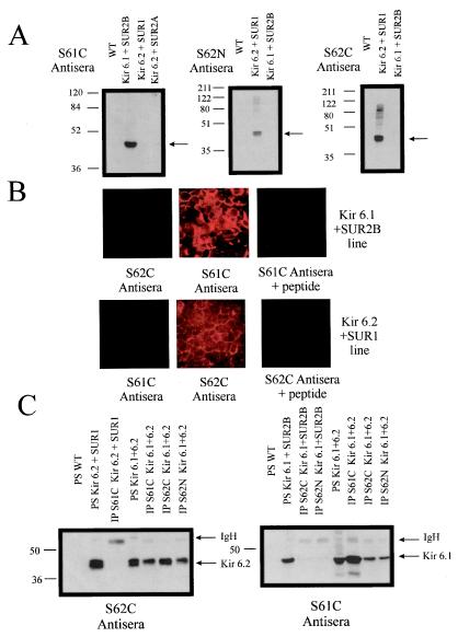
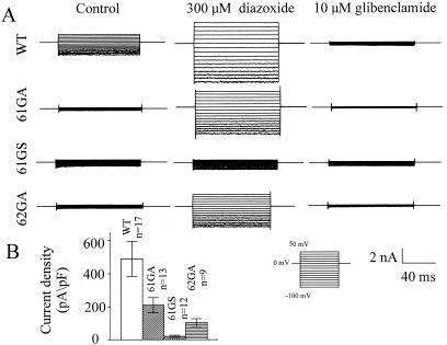
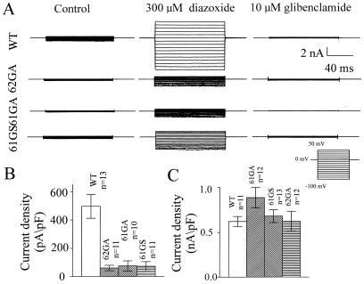
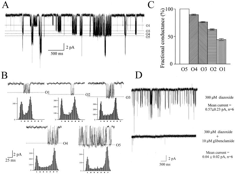
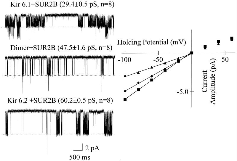
ATP-sensitive potassium channels are an octomeric complex of four pore-forming subunits of the Kir 6.0 family and four sulfonylurea receptors. The Kir 6.0 family consists of two known members, Kir 6.1 and Kir 6.2, with distinct functional properties. The tetrameric structure of the pore-forming domain leads to the possibility that mixed heteromultimers may form. In this study, we examine this by using biochemical and electrophysiological techniques after heterologous expression of these subunits in HEK293 cells. After the coexpression of Kir 6.1 and Kir 6.2, Kir 6.1 can be coimmunoprecipitated with isoform-specific Kir 6.2 antisera and vice versa. Coexpression of SUR2B and Kir 6.2 with Kir 6.1 dominant negatives at a 1:1 expression ratio and vice versa led to a potent suppression of current. Kir 6.1, and Kir 6.2 dominant negative mutants were without effect on an inwardly rectifying potassium channel from a different family, Kir 2.1. Single-channel analysis, after coexpression of SUR2B, Kir 6.1, and Kir 6.2, revealed the existence of five distinct populations with differing single-channel current amplitudes. All channel populations were inhibited by glibenclamide. A dimeric Kir 6.1-Kir 6.2 construct expressed with SUR2B had a single-channel conductance intermediate between that of either Kir 6.2 or Kir 6.1 expressed with SUR2B. In conclusion, Kir 6.1 and Kir 6.2 readily coassemble to produce functional channels, and such phenomena may contribute to the diversity of nucleotide-regulated potassium currents seen in native tissues.


Figures

Figure 1
Figure 1
Characterization of antisera by Western blotting and immunofluorescence and coimmunoprecipitation of Kir 6.1 and Kir 6.2. (A) Western blots characterizing Kir 6.0 subtype antisera. Samples were run on a 10% (S61C probed blot) or a 12% (S62C/N probed blot) denaturing polyacrylamide gel. The amount of each sample loaded was 3.5 μg, and all blots were exposed to film for 30 s. The position of the Kir 6.0 subtype is indicated by the arrows, and the molecular weight markers are shown alongside the blots. Kir 6.1 migrates as an approximately 48-kDa protein and Kir 6.2 migrates as an approximately 44-kDa protein. The lanes labeled WT denote untransfected HEK293 cells. (B) Characterization of immunofluorescence against the Kir 6.1 + SUR2B and the Kir 6.2 + SUR1 lines. Immunofluorescent staining and analysis was as described in Methods. The subtype specificity of immunostaining and competition by preincubation with the immunogenic peptide (1 mg/ml) can be observed. (C) On the Left, a Western blot (from a 12% denaturing polyacrylamide gel) probed with the S62C antisera. From Left to Right, the first three lanes show that Kir 6.2 is present in the SUR1 + Kir 6.2 cell line (PS, protein sample) and that the Kir 6.1C antiserum does not immunoprecipitate Kir 6.2. The next four lanes show immunoprecipitation studies done on a stable line coexpressing Kir 6.1 and Kir 6.2. As expected, Kir 6.2 is immunoprecipitated by both S62C and S62N, but Kir 6.2 is also coimmunoprecipitated by the S61C antiserum. On the Right, a Western blot (from a 10% denaturing polyacrylamide gel) probed with the S61C antisera. From Left to Right, the first four lanes show that Kir 6.1 is present in the SUR2B + Kir 6.1 cell line (PS, denatured protein sample homogenate taken before IP) and that the S62N and S62C antiserum do not immunoprecipitate Kir 6.1. The next four lanes show the samples of homogenate and immunoprecipitation reactions from the Kir 6.1 + Kir 6.2 stable line, and they show that Kir 6.1 is immunoprecipitated by the S61C antiserum and also coimmunoprecipitated by the S62C and S62N antisera (same samples as exposed to the S62C antisera on the Left). Both Western blots were exposed to film for 30 s. The positions of Kir 6.1, Kir 6.2, and the Ig heavy chain (IgH, only a small amount present because of the covalent linkage to protein A) are indicated by the black arrows. Molecular weight markers are shown alongside the blots.
Figure 2
Figure 2
Dominant negative effects of 61GA, 61GS, and 62GA on SUR2B + Kir 6.1 current expression. (A) Whole-cell current traces recorded from cells transiently transfected with SUR2B + Kir 6.1 (WT), SUR2B + Kir 6.1 + 61GA (61GA), SUR2B + Kir 6.1 + 61GS (61GS), or SUR2B + Kir 6.1 + 62GA (62GA) under the control conditions (5 min after breakin), in the presence of diazoxide (300 μM) and glibenclamide (10 μM), respectively. The pipette solution (see Methods) was supplemented with 0.5 mM GDP. (B) Summarized data for the currents measured at −100 mV. P < 0.01 one-way ANOVA.
Figure 3
Figure 3
Dominant negative effects of 62GA, 61GA, and 61GS on SUR2B + Kir 6.2 current. (A) Whole-cell current traces recorded from cells transiently transfected with SUR2B + Kir 6.2 (WT), SUR2B + Kir 6.2 + 62GA (62GA), SUR2B + Kir 6.2 + 61GA (61GA), or SUR2B + Kir 6.2 + 61GS (61GS) under the control conditions (5 min after breakin), in the presence of diazoxide (300 μM) and glibenclamide (10 μM), respectively. The pipette solution (see Methods) was supplemented with 3.0 mM ATP. (B) Summarized data for the currents measured at −100 mV. P < 0.01, one-way ANOVA. (C) Mean measured currents at −100 mV recorded from cells transiently transfected with Kir 2.1–His6 and cotransfected with Kir 2.1–His6 and the indicated Kir 6.0 constructs (5 min after breakin).
Figure 4
Figure 4
Five different conductance channels in cells coexpressing SUR2B, Kir 6.1, and Kir 6.2. (A) Single-channel current trace (cell-attached configuration) recorded at −100 mV in the presence of 300 μM diazoxide from Kir 6.1 + Kir 6.2 stable cell line transiently transfected with SUR2B. (B) Representative recordings of five single channel unitary currents (O1–O5) at −100 mV in the presence of 300 μM diazoxide and their corresponding amplitude histograms. (C) Bar chart showing fractional current amplitudes normalized to the O5 channel conductance. (D) Pharmacological sensitivity of single-channel currents recorded from Kir 6.1 + Kir 6.2 stable cell line transiently transfected with SUR2B. The current amplitude was measured as a mean current over 20 s recorded at −100 mV and significantly reduced in the presence of 10 μM glibenclamide (P < 0.05, paired t test).
Figure 5
Figure 5
Single-channel conductances of SUR2B + Kir 6.1, SUR2B + Kir 6.1–Kir 6.2 dimer and SUR2B + Kir 6.2 currents. On the Left, single-channel current traces (cell-attached configuration) recorded at −100 mV from SUR2B + Kir 6.1and SUR2B + Kir 6.2 stable cell lines and cells transiently transfected with SUR2B + Kir 6.1—Kir 6.2 dimer in the presence of 300 μM diazoxide. The single-channel conductances shown are significantly different (P < 0.01, one-way ANOVA). On the Right, the current–voltage relationships of SUR2B + Kir 6.1 (▴), SUR2B + Kir 6.1–Kir 6.2 dimer (⧫), and SUR2B + Kir6.2 (■) currents.

Similar articles

Cited by

References

    1. Jan L Y, Jan Y N. Annu Rev Neurosci. 1997;20:91–123. - PubMed
    1. Nichols C G, Lopatin A N. Annu Rev Physiol. 1997;59:171–191. - PubMed
    1. Li M, Jan Y N, Jan L Y. Science. 1992;257:1225–1230. - PubMed
    1. Isacoff E Y, Jan Y N, Jan L Y. Nature (London) 1990;345:530–534. - PubMed
    1. Sheng M, Liao Y J, Jan Y N, Jan L Y. Nature (London) 1993;365:72–75. - PubMed

Publication types

MeSH terms

LinkOut - more resources