

Macrophage are the principal reservoir and sustain high virus loads in rhesus macaques after the depletion of CD4+ T cells by a highly pathogenic simian immunodeficiency virus/HIV type 1 chimera (SHIV): Implications for HIV-1 infections of humans
- PMID: 11136236
- PMCID: PMC14644
- DOI: 10.1073/pnas.98.2.658
Macrophage are the principal reservoir and sustain high virus loads in rhesus macaques after the depletion of CD4+ T cells by a highly pathogenic simian immunodeficiency virus/HIV type 1 chimera (SHIV): Implications for HIV-1 infections of humans
Abstract
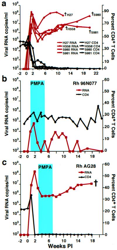
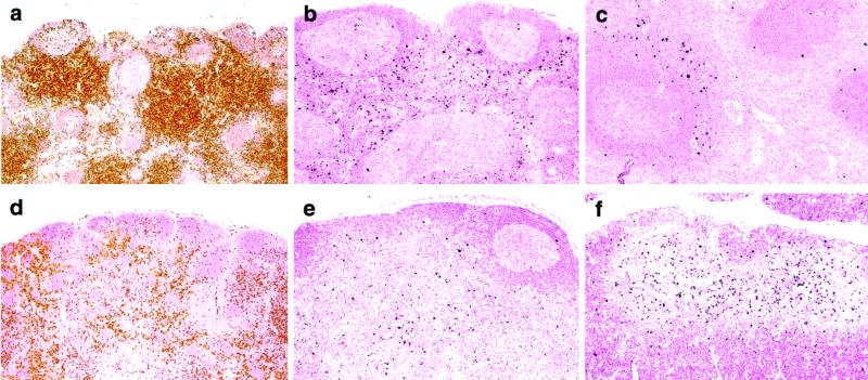
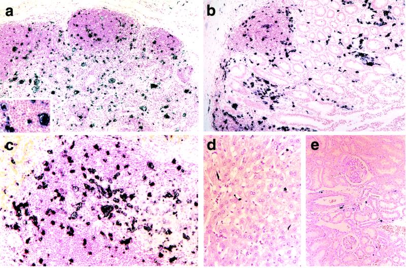
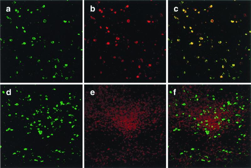
The highly pathogenic simian immunodeficiency virus/HIV type 1 (SHIV) chimeric virus SHIV(DH12R) induces a systemic depletion of CD4(+) T lymphocytes in rhesus monkeys during the initial 3-4 weeks of infection. Nonetheless, high levels of viral RNA production continue unabated for an additional 2-5 months. In situ hybridization and immunohistochemical analyses revealed that tissue macrophage in the lymph nodes, spleen, gastrointestinal tract, liver, and kidney sustain high plasma virus loads in the absence of CD4(+) T cells. Quantitative confocal immunofluorescence analysis indicated that greater than 95% of the virus-producing cells in these tissues are macrophage and less than 2% are T lymphocytes. Interestingly, the administration of a potent reverse transcriptase inhibitor blocked virus production during the early T cell phase but not during the later macrophage phase of the SHIV(DH12R) infection. When interpreted in the context of HIV-1 infections, these results implicate tissue macrophage as an important reservoir of virus in vivo. They become infected during the acute infection, gradually increase in number over time, and can be a major contributor to total body virus burden during the symptomatic phase of the human infection.
Figures
References
-
- Gulick R M, Mellors J W, Havlir D, Eron J J, Gonzalez C, McMahon D, Richman D D, Valentine F T, Jonas L, Meibohm A, et al. N Engl J Med. 1997;337:734–739. - PubMed
-
- Palella F J, Jr, Delaney K M, Moorman A C, Loveless M O, Fuhrer J, Satten G A, Aschman D J, Holmberg S D. N Engl J Med. 1998;338:853–860. - PubMed
-
- Natarajan V, Bosche M, Metcalf J A, Ward D J, Lane H C, Kovacs J A. Lancet. 1999;353:119–120. - PubMed
-
- Furtado M R, Callaway D S, Phair J P, Kunstman K J, Stanton J L, Macken C A, Perelson A S, Wolinsky S M. N Engl J Med. 1999;340:1614–1622. - PubMed
-
- Zhang L, Ramratnam B, Tenner-Racz K, He Y, Vesanen M, Lewin S, Talal A, Racz P, Perelson A S, Korber B T, et al. N Engl J Med. 1999;340:1605–1613. - PubMed
MeSH terms
Substances
LinkOut - more resources
Full Text Sources
Other Literature Sources
Medical
Research Materials
