. 2001 Jan 2;98(1):105-12.
doi: 10.1073/pnas.98.1.105.

An amino acid code for protein folding

Affiliations

An amino acid code for protein folding

J Rumbley et al. Proc Natl Acad Sci U S A. .

Abstract

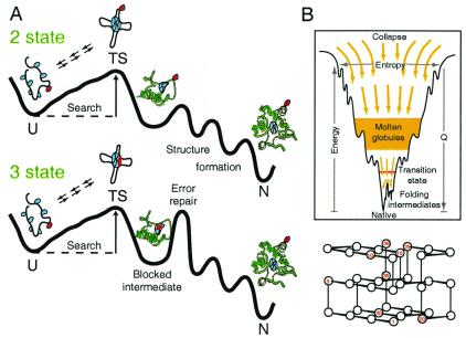
Direct structural information obtained for many proteins supports the following conclusions. The amino acid sequences of proteins can stabilize not only the final native state but also a small set of discrete partially folded native-like intermediates. Intermediates are formed in steps that use as units the cooperative secondary structural elements of the native protein. Earlier intermediates guide the addition of subsequent units in a process of sequential stabilization mediated by native-like tertiary interactions. The resulting stepwise self-assembly process automatically constructs a folding pathway, whether linear or branched. These conclusions are drawn mainly from hydrogen exchange-based methods, which can depict the structure of infinitesimally populated folding intermediates at equilibrium and kinetic intermediates with subsecond lifetimes. Other kinetic studies show that the polypeptide chain enters the folding pathway after an initial free-energy-uphill conformational search. The search culminates by finding a native-like topology that can support forward (native-like) folding in a free-energy-downhill manner. This condition automatically defines an initial transition state, the search for which sets the maximum possible (two-state) folding rate. It also extends the sequential stabilization strategy, which depends on a native-like context, to the first step in the folding process. Thus the native structure naturally generates its own folding pathway. The same amino acid code that translates into the final equilibrium native structure-by virtue of propensities, patterning, secondary structural cueing, and tertiary context-also produces its kinetic accessibility.


Figures

Figure 1
Figure 1
Alternative folding paradigms. (A) Classical pathways. Following Sosnick et al. (62), the profiles shown suggest a basis for apparent two-state folding (Upper) due to an initial rate-limiting barrier, even though intermediates are present; three-state folding (Lower) due to the insertion of an error repair barrier (101, 107); and heterogeneous folding (Upper and Lower) due to the chance nature of misfolding errors. The molecular cartoons are meant to suggest a lengthy energetically uphill conformational search for a set of interactions that can pin the chain into some native-like transition-state (TS) topology, the subsequent downhill folding that marks the success of the search, and the chance incorrect placement of some group(s) (red) that can block folding and impose a time-consuming error repair process. This picture is formalized in Eq. 2. (B) A folding funnel (adapted from ref. 13) and a typical on-lattice folding model (adapted from ref. 108).
Figure 2
Figure 2
HX labeling data. (A and B) The fraction of H labeling obtained at particular residue amides in Cyt c by a brief high-pH labeling pulse after increasing times of folding (36). The data indicate the early formation of an intermediate with the N- and C-terminal helices formed, the delayed formation of the rest of the protein, and heterogeneous folding in which different fractions of the protein population fold at different rates. (C) Native-like HX protection pattern for the Cyt c molten globule destabilized at acid pH (122). Protection factors are corrected (123, 124). Color coding relates to Fig. 3D. Residues 14, 15, and 18 (gray) are protected within the small covalent heme loop, even in the unfolded state (see Fig. 3A).
Figure 3
Figure 3
Native-state HX results for Cyt c [pDr 7, 30°C (48)]. (A) Residues in the N-terminal helix. (B) Residues in the 60s helix. Local fluctuational HX pathways, dominant at low denaturant (m ≈ 0), are superseded by larger unfolding reactions when denaturant is increased. (C) A summary crossover curve showing how the four different unfolded states change in free energy, relative to N and to each other, with GdmCl concentration. The dashed line suggests the formation of a partially unfolded molten globule because of some selective destabilization of an unfolding unit. (D) Cyt c structure color coded in order of descending stabilization free energy (blue to green to yellow to red) to show the cooperative units indicated by native-state HX. Also shown are the peripheral histidines that can misligate to the heme iron at the Met-80-S site and trap the green loop out of place, imposing an error correction barrier that causes slow multistate folding with accumulation of the N/C helix intermediate.
Figure 4
Figure 4
Native contact diagram for the blue, green, yellow, and red folding units in equine Cyt c. Connected residues have atom–atom contacts within 4 Å in the native structure (125) without distinction between polar or apolar character. Extensive contacts connect the two blue segments (Top), the blue segments with the rest of the protein (Middle), and the green segments with the rest of the protein (excluding blue) (Bottom). Folding by sequential stabilization in the native context would explain the concerted initial formation of the N- and C-terminal helices, the subsequent apparently concerted formation of both green elements, and the later folding of the yellow and red elements.

Similar articles

Cited by

References

    1. Anfinsen C B. Science. 1973;181:223–230. - PubMed
    1. Creighton T E. Adv Biophys. 1984;18:1–20. - PubMed
    1. Kim P S, Baldwin R L. Annu Rev Biochem. 1990;59:631–660. - PubMed
    1. Baldwin R L. Nat Struct Biol. 1999;6:814–817. - PubMed
    1. Fersht A R, Serrano L. Curr Opin Struct Biol. 1993;3:75–83.

Publication types

LinkOut - more resources