. 2001 Jan 1;193(1):61-71.
doi: 10.1084/jem.193.1.61.

A regulatory role for Src homology 2 domain-containing inositol 5'-phosphatase (SHIP) in phagocytosis mediated by Fc gamma receptors and complement receptor 3 (alpha(M)beta(2); CD11b/CD18)

Affiliations

A regulatory role for Src homology 2 domain-containing inositol 5'-phosphatase (SHIP) in phagocytosis mediated by Fc gamma receptors and complement receptor 3 (alpha(M)beta(2); CD11b/CD18)

D Cox et al. J Exp Med. .

Abstract

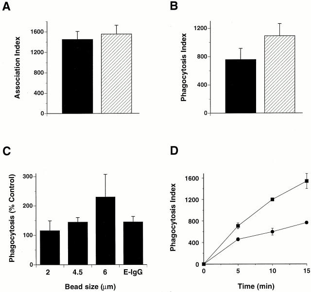
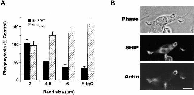
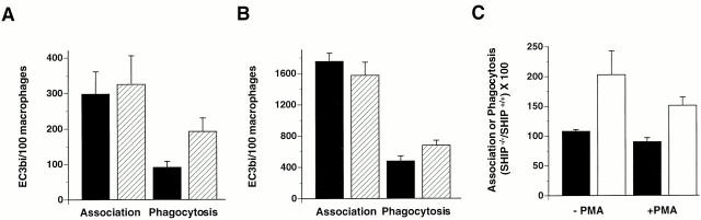
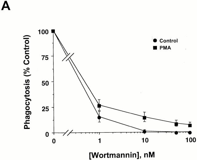
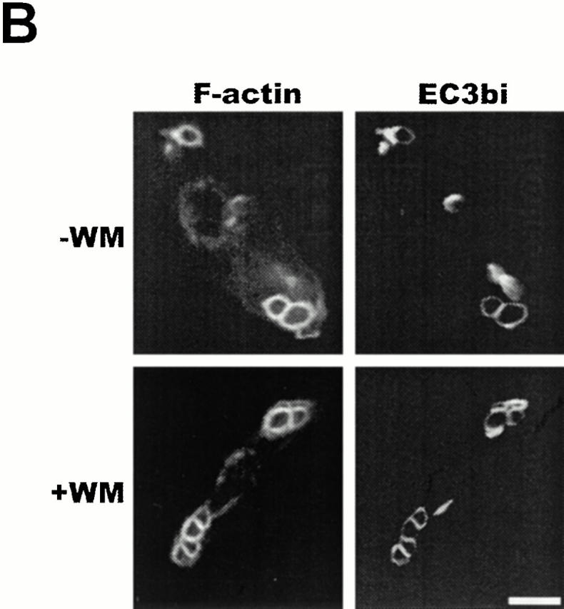
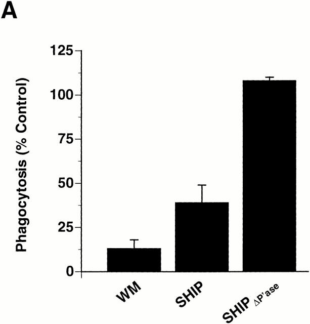
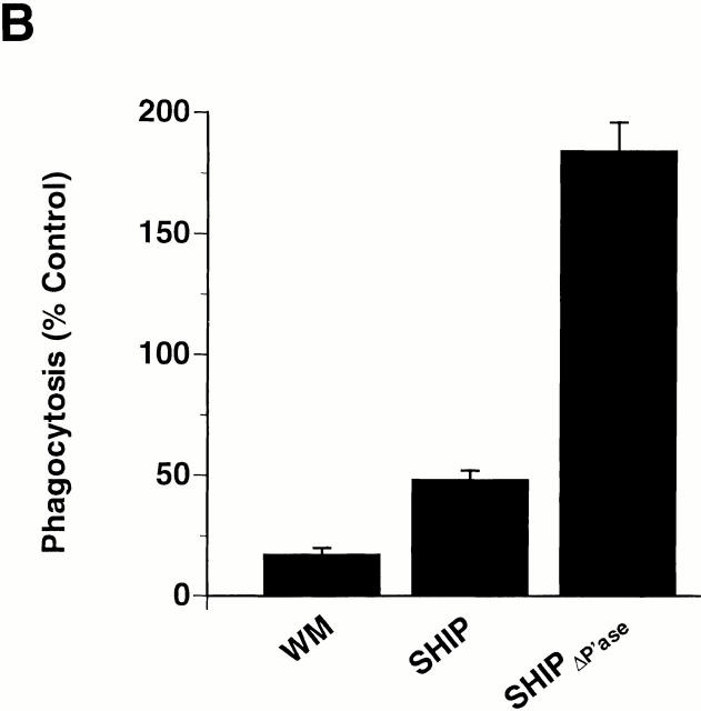
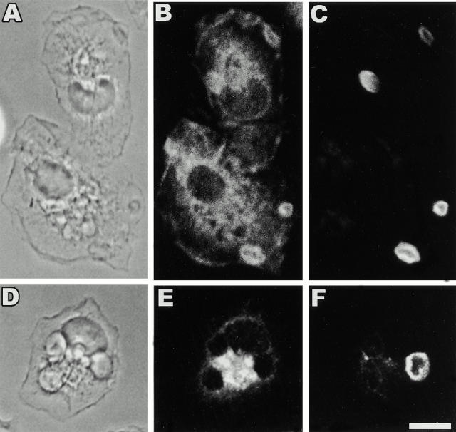
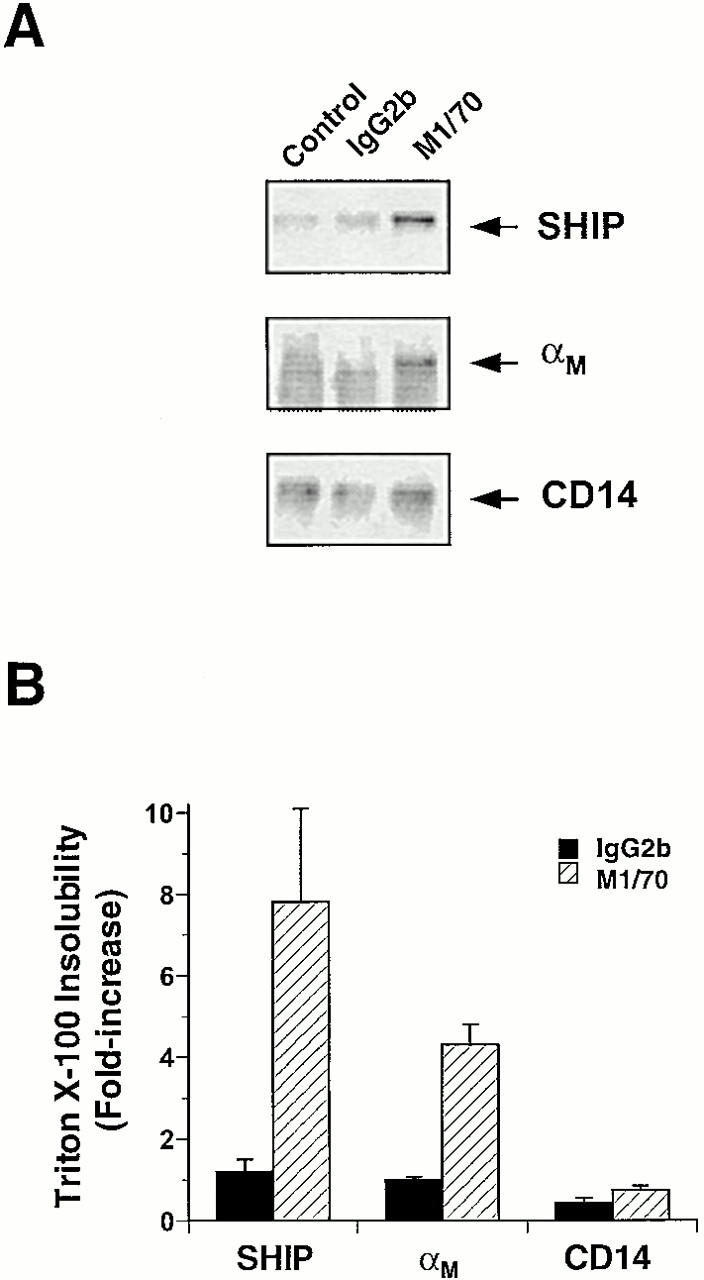
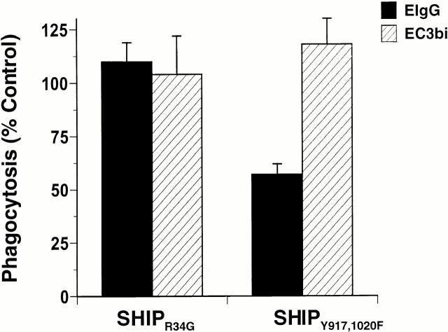
The Src homology 2 domain-containing inositol 5'-phosphatase (SHIP) is recruited to immunoreceptor tyrosine-based inhibition motif (ITIM)-containing proteins, thereby suppressing phosphatidylinositol 3-kinase (PI 3-kinase)-dependent pathways. The role of SHIP in phagocytosis, a PI 3-kinase-dependent pathway, is unknown. Overexpression of SHIP in macrophages led to an inhibition of phagocytosis mediated by receptors for the Fc portion of IgG (Fc gamma Rs). In contrast, macrophages expressing catalytically inactive SHIP or lacking SHIP expression demonstrated enhanced phagocytosis. To determine whether SHIP regulates phagocytosis mediated by receptors that are not known to recruit ITIMs, we determined the effect of SHIP expression on complement receptor 3 (CR3; CD11b/CD18; alpha(M)beta(2))-dependent phagocytosis. Macrophages overexpressing SHIP demonstrated impaired CR3-mediated phagocytosis, whereas macrophages expressing catalytically inactive SHIP demonstrated enhanced phagocytosis. CR3-mediated phagocytosis in macrophages derived from SHIP(-/-) mice was up to 2.5 times as efficient as that observed in macrophages derived from littermate controls. SHIP was localized to Fc gamma R- and CR3-containing phagocytic cups and was recruited to the cytoskeleton upon clustering of CR3. In a transfected COS cell model of activation-independent CR3-mediated phagocytosis, catalytically active but not inactive SHIP also inhibited phagocytosis. We conclude that PI 3-kinase(s) and SHIP regulate multiple forms of phagocytosis and that endogenous SHIP plays a role in modulating beta(2) integrin outside-in signaling.


Figures

Figure 1
Figure 1
Macrophages lacking SHIP demonstrate enhanced FcγR-mediated phagocytosis. (A and B) Thio-macrophages derived from SHIP+/+ (black bars) and SHIP−/− (hatched bars) mice were challenged with EIgG for 15 min at 37°C as described in Materials and Methods. Association and phagocytic indices are shown. Data represent mean ± SEM, n = 5 (association) and 8 (phagocytosis). Differences between phagocytosis, but not binding, in SHIP+/+ and SHIP−/− macrophages were significant (P < 0.05). (C) Phagocytosis of SHIP−/− macrophages cells ingesting EIgG or IgG-coated latex beads expressed as a percentage of phagocytosis in SHIP+/+ macrophages. Phagocytic indices for 2-, 4.5-, and 6-μm beads, and EIgG, in SHIP+/+ macrophages were 540 ± 18, 1,284 ± 105, 140 ± 22, and 761 ± 55, respectively. Data represent mean ± SEM, n = 4–8. Differences between phagocytosis of 4.5- and 6-μm beads, and EIgG, but not 2-μm beads, in SHIP+/+ and SHIP−/− macrophages were significant (P < 0.05). (D) Time course of synchronized phagocytosis of EIgG in macrophages derived from SHIP+/+ (•) and SHIP /− (▪) mice. Macrophages were incubated with EIgG at 4°C to allow binding, but not ingestion, and further incubated for the indicated times at 37°C to allow for ingestion. Data represent mean ± SEM, n = 3.
Figure 2
Figure 2
Endogenously expressed SHIP modulates FcγR-mediated phagocytosis. (A) Adherent RAW LR5 cells transfected with Myc-tagged wild-type SHIP (WT) or phosphatase-deficient SHIP (SHIPΔPase) were challenged with EIgG or IgG-coated beads for 30 min at 37°C as described in Materials and Methods. Phagocytosis of cells overexpressing SHIP (black bars) or expressing SHIPΔPase (hatched bars) is reported as a percentage of nonexpressing controls. Phagocytic indices for 2-, 4.5-, and 6-μm beads, and EIgG, in control macrophages were 652 ± 120, 423 ± 73, 261 ± 30, and 301 ± 34, respectively. Data represent mean ± SEM, n = 3–6. Differences between phagocytosis of macrophages expressing either wild-type SHIP or SHIPΔPase, and nonexpressing controls were significant (P values of 0.03 or less) for all but the 2-μm beads. (B) Adherent thio-macrophages obtained from SHIP+/+ mice were incubated with EIgG2a for 7 min at 37°C. After fixation, cells were stained for the presence of SHIP and EIgG2a as described in Materials and Methods. Bar, 10 μm.
Figure 3
Figure 3
Lack of SHIP expression is associated with enhanced CR3-mediated phagocytosis. Adherent thio-macrophages obtained from SHIP+/+ (black bars) or SHIP−/− (hatched bars) mice were incubated with EC3bi for 30 min at 37°C. The association and phagocytosis indices were determined for cells incubated in the absence (A) and/or presence (B) of 100 nM PMA. Phagocytic indices in SHIP+/+ macrophages were 91 ± 17 in the absence of PMA and 479 ± 65 in the presence of PMA. (C) Phagocytosis of EC3bi expressed as a percentage, SHIP−/− versus SHIP+/+. Data represent mean ± SEM, n = 5–8. Differences between phagocytosis, but not binding, in SHIP+/+ and SHIP−/− macrophages were significant (P < 0.05).
Figure 4
Figure 4
CR3-mediated phagocytosis, but not actin assembly, is dependent on PI 3-kinase. (A) Adherent thio-macrophages were preincubated for 15 min with the indicated concentrations of WM followed by the addition of EC3bi in the presence or absence of 100 nM PMA for 30 min at 37°C. Phagocytic indices were 84 ± 9 in the absence of PMA and 472 ± 125 in the presence of PMA. Phagocytosis of cells incubated with WM is expressed as the percentage of phagocytosis performed in the absence of WM. Data represent mean ± SEM, n = 3. (B) Adherent thio-macrophages were preincubated without (−WM) or with (+WM) 100 nM WM followed by further incubation with EC3bi for 12 min at 37°C. After fixation, cells were stained for F-actin with rhodamine-phalloidin and for EC3bi with anti-EIgG followed by FITC-anti–rabbit IgG. Bar, 10 μm.
Figure 4
Figure 4
CR3-mediated phagocytosis, but not actin assembly, is dependent on PI 3-kinase. (A) Adherent thio-macrophages were preincubated for 15 min with the indicated concentrations of WM followed by the addition of EC3bi in the presence or absence of 100 nM PMA for 30 min at 37°C. Phagocytic indices were 84 ± 9 in the absence of PMA and 472 ± 125 in the presence of PMA. Phagocytosis of cells incubated with WM is expressed as the percentage of phagocytosis performed in the absence of WM. Data represent mean ± SEM, n = 3. (B) Adherent thio-macrophages were preincubated without (−WM) or with (+WM) 100 nM WM followed by further incubation with EC3bi for 12 min at 37°C. After fixation, cells were stained for F-actin with rhodamine-phalloidin and for EC3bi with anti-EIgG followed by FITC-anti–rabbit IgG. Bar, 10 μm.
Figure 5
Figure 5
Effect of SHIP or SHIPΔPase expression on CR3-mediated phagocytosis in COS cells and macrophages. (A) COS cells transfected with the αM and β2 subunits of CR3 and coexpressing the indicated constructs were challenged with EC3bi for 30 min at 37°C in the presence or absence of 100 nM WM. The phagocytosis index of COS cells expressing CR3 alone, in the absence of WM (controls) was 651 ± 108. Phagocytosis was determined as the percentage of control. Data represent mean ± SEM, n = 3. Differences in phagocytosis between cells either incubated in WM or expressing wild-type SHIP, and controls were significant (P < 0.05). (B) RAW LR5 cells expressing the indicated constructs were challenged with EC3bi and 100 nM PMA and incubated in the absence or presence of 100 nM WM for 30 min at 37°C. The phagocytosis index of nonexpressing cells in the absence of WM (controls) was 55 ± 10. Phagocytosis was determined as the percentage of controls. Data represent mean ± SEM, n = 5. Differences in phagocytosis between cells either incubated in WM, or expressing wild-type SHIP or SHIPΔPase, and controls were significant (P < 0.05).
Figure 5
Figure 5
Effect of SHIP or SHIPΔPase expression on CR3-mediated phagocytosis in COS cells and macrophages. (A) COS cells transfected with the αM and β2 subunits of CR3 and coexpressing the indicated constructs were challenged with EC3bi for 30 min at 37°C in the presence or absence of 100 nM WM. The phagocytosis index of COS cells expressing CR3 alone, in the absence of WM (controls) was 651 ± 108. Phagocytosis was determined as the percentage of control. Data represent mean ± SEM, n = 3. Differences in phagocytosis between cells either incubated in WM or expressing wild-type SHIP, and controls were significant (P < 0.05). (B) RAW LR5 cells expressing the indicated constructs were challenged with EC3bi and 100 nM PMA and incubated in the absence or presence of 100 nM WM for 30 min at 37°C. The phagocytosis index of nonexpressing cells in the absence of WM (controls) was 55 ± 10. Phagocytosis was determined as the percentage of controls. Data represent mean ± SEM, n = 5. Differences in phagocytosis between cells either incubated in WM, or expressing wild-type SHIP or SHIPΔPase, and controls were significant (P < 0.05).
Figure 6
Figure 6
SHIP, but not c-fms, localizes to CR3-mediated phagocytic cups. Adherent thio-macrophages obtained from SHIP+/+ mice were incubated with EC3bi, as described in Materials and Methods, for 12 min at 37°C. After fixation, cells were stained for the presence of SHIP or c-fms (negative control), and C3bi as described in Materials and Methods. (A and D) Phase–contrast; (B) anti-SHIP; (E) anti–c-fms; (C and F) anti-C3. Bar, 10 μm.
Figure 7
Figure 7
Incorporation of SHIP and αM into the Triton X-100–insoluble cytoskeleton after CR3 clustering. Triton X-100–insoluble fractions were isolated from control cells or from cells incubated with mAb M1/70 or an isotype-matched control mAb and F(ab′)2 fragments of goat anti–rat IgG as described in Materials and Methods. (A) Representative immunoblot of cytoskeletons probed with the indicated antibodies. (B) Fold increase in cytoskeletal incorporation of the indicated proteins. Data represent mean ± SEM, n = 3.
Figure 8
Figure 8
Differential requirements for the NPXY motifs of SHIP in inhibition of FcγR- and CR3-mediated phagocytosis. RAW LR5 cells transfected with the indicated constructs were challenged with either EIgG or EC3bi in the presence of 100 nM PMA, and incubated for 30 min at 37°C. Phagocytic indices were 530 ± 49 for EIgG and 104 ± 17 for EC3bi. Phagocytosis of cells expressing the indicated constructs is reported as the percentage of nonexpressing controls. Data represent mean ± SEM, n = 3. Difference in phagocytosis of EIgG by cells expressing SHIPY917,1020F and controls was significant (P < 0.05).

References

    1. Cox D., Tseng C.-C., Bjekic G., Greenberg S. A requirement for phosphatidylinositol 3-kinase in pseudopod extension. J. Biol. Chem. 1999;274:1240–1247. - PubMed
    1. Araki N., Johnson M.T., Swanson J.A. A role for phosphoinositide 3-kinase in the completion of macropinocytosis and phagocytosis by macrophages. J. Cell Biol. 1996;135:1249–1260. - PMC - PubMed
    1. Huber M., Helgason C.D., Damen J.E., Scheid M., Duronio V., Liu L., Ware M.D., Humphries R.K., Krystal G. The role of SHIP in growth factor induced signalling. Prog. Biophys. Mol. Biol. 1999;71:423–434. - PubMed
    1. Rohrschneider L.R., Fuller J.F., Wolf I., Liu Y., Lucas D.M. Structure, function, and biology of SHIP proteins. Genes Dev. 2000;14:505–520. - PubMed
    1. Ono M., Okada H., Bolland S., Yanagi S., Kurosaki T., Ravetch J.V. Deletion of SHIP or SHP-1 reveals two distinct pathways for inhibitory signaling. Cell. 1997;90:293–301. - PubMed

Publication types

Substances